. 2022 Jan;72(1):136-148.
doi: 10.1007/s12031-021-01916-5. Epub 2021 Sep 26.

MiR-615 Agomir Encapsulated in Pluronic F-127 Alleviates Neuron Damage and Facilitates Function Recovery After Brachial Plexus Avulsion

Affiliations

MiR-615 Agomir Encapsulated in Pluronic F-127 Alleviates Neuron Damage and Facilitates Function Recovery After Brachial Plexus Avulsion

Kangzhen Chen et al. J Mol Neurosci. 2022 Jan.

Abstract

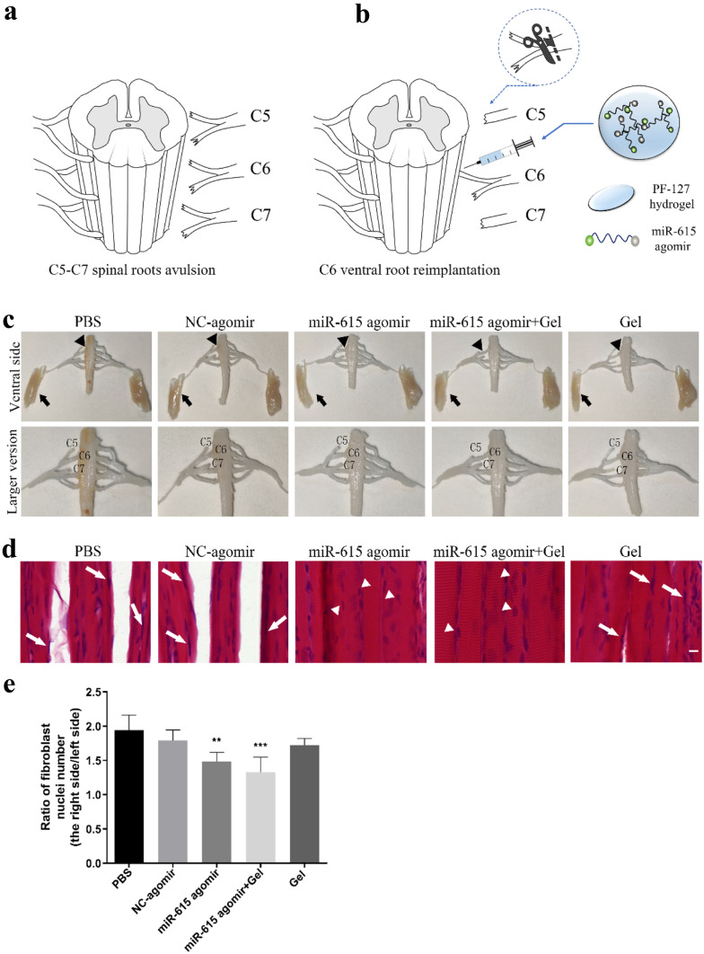
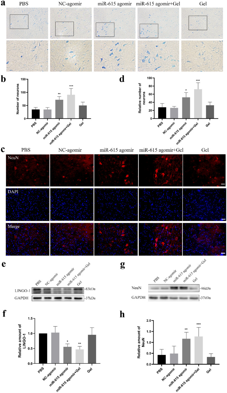
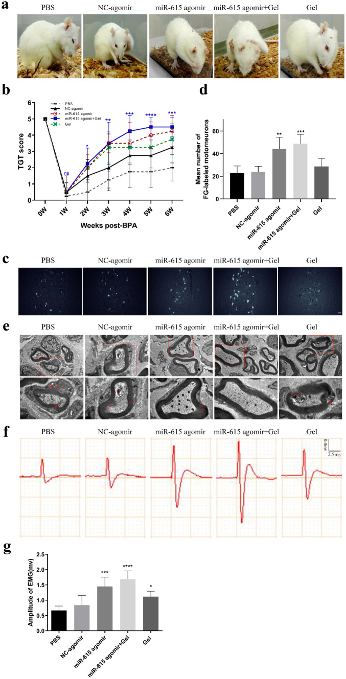
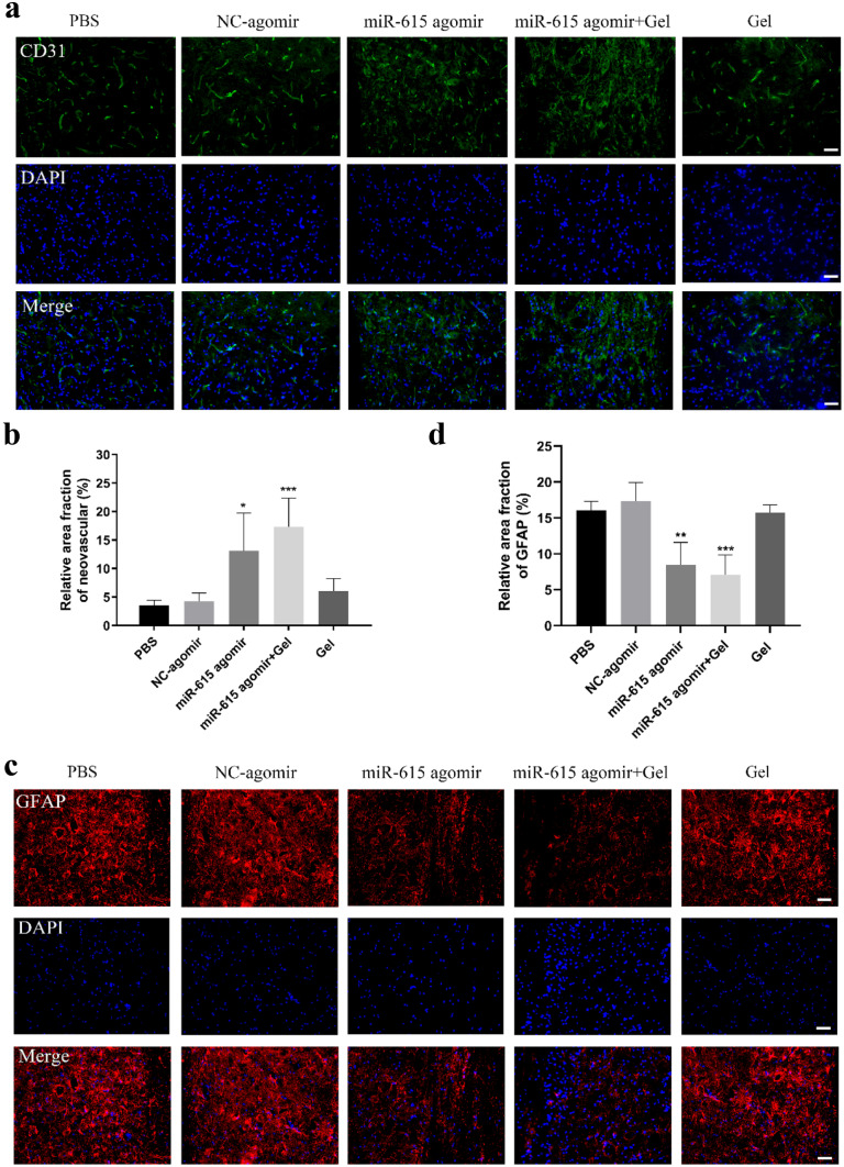
Brachial plexus avulsion (BPA) is a devastating traumatic peripheral nerve injury complicated with paralysis of the upper extremity. We previously reported that leucine-rich repeat and immunoglobulin-like domain-containing NOGO receptor-interacting protein 1 (LINGO-1) has a potent role in inhibiting neuron survival and axonal regeneration after the central nervous system (CNS) damage and miR-615 is a potential microRNA (miRNA) negatively regulated LINGO-1. However, the effect of miR-615 in BPA remains to be elucidated. Accumulating evidence indicates that pluronic F-127 (PF-127) hydrogel could serve as a promising vehicle for miRNA encapsulation. Thus, to further explore the potential role of hydrogel-miR-615 in BPA-reimplantation, the present study established the BPA rat model and injected miR-615 agomir encapsulated by PF-127 hydrogel into the reimplantation site using a microsyringe. In this study, results indicated that hydrogel-miR-615 agomir effectively alleviated motoneuron loss by LINGO-1 inhibition, promoted musculocutaneous nerve regeneration and myelination, reduced astrocytes activation, promoted angiogenesis and attenuated peripheral amyotrophy, leading to improved motor functional rehabilitation of the upper extremity. In conclusion, our findings demonstrate that miR-615-loaded PF-127 hydrogel may represent a novel therapeutic strategy for BPA treatment.

Keywords: Brachial plexus avulsion (BPA); MiR-615; Motoneuron; Pluronic F-127(PF-127).


Conflict of interest statement

No conflict of interest exists in the submission of this manuscript, and this manuscript is approved by all authors listed for publication.

The authors declare no competing interests.

Figures

Fig. 1
Fig. 1
Co-transplantation of miR-615 agomir with gel alleviated muscle atrophy after avulsion/reimplantation. a Right-sided avulsion of C5–C7 spinal nerve roots and reimplantation of C6 ventral root into the surface of the corresponding spinal segment. b The injector indicates the reimplanted site and the miR-615 agomir and pluronic F-127 hydrogel injection site. c Representative pictures of gross anatomical specimen of rats with BPA reimplantation from different groups. Reimplanted C6 ventral roots (black triangle) tightly connected to the original avulsion site. On the contrary, two obvious defects were observed between C5 and C7 nerve roots and the corresponding spinal segments. Compared with the healthy side, atrophied biceps were common in the affected side (black arrows). d Longitudinal sections of the right affected biceps by hematoxylin and eosin staining. In PBS and NC-agomir groups, shrunken sarcoplasm and large fibroblast nucleus (white arrow) were common in atrophied biceps. Instead, miR-615 agomir and miR-615 agomir+gel group showed less fibrosis, demonstrated by fewer fibroblast nucleus and a great many of clear myocyte nuclei (white triangle). e The level of fibrosis was decided by the ratio of fibroblast nuclei number in ipsilateral to contralateral biceps. Data are presented as the mean ± SD (one-way analysis of variance followed by the least significant difference post hoc test). **p < 0.01, ***p < 0.001, vs. PBS group. Scale bar (C): 100 μm
Fig. 2
Fig. 2
Co-transplantation of miR-615 agomir with gel stimulated axonal regeneration and reduced neuronal death in affected spinal segments after avulsion/reimplantation. a, b Representative micrographs and quantitative analysis of the survival neurons around the avulsed epicenter by Nissl’s staining at low and high magnification, respectively, indicating a significant increasement of neurons in miR-615 agomir+gel rats. c Immunofluorescence photos of neurons around the avulsed site in the ipsilateral C6 segment. d Relative number of neurons were measured and counted. e, f Western blot assay and relative quantification of LINGO-1 protein demonstrated dramatic downregulation of LINGO-1 in miR-615 agomir and miR-615 agomir+gel group. The results illustrated that miR-615 negatively regulated LINGO-1. GAPDH was used as an internal control. g, h Western blot assay and relative quantification of NeuN, a neuron-specific protein, demonstrated dramatic upregulation of NeuN in miR-615 agomir and miR-615 agomir+gel group. GAPDH was used as an internal control. Data are presented as the mean ± SD (one-way analysis of variance followed by the least significant difference post hoc test). *p < 0.05, **p < 0.01, ***p < 0.001, vs. PBS group. Scale bar (a—upper row) = 500 μm, scale bar (b—lower row) = 250 μm, Scale bar (c) = 200 μm
Fig. 3
Fig. 3
Co-transplantation of miR-615 agomir with gel improved the motor functional restoration of monoplegia right upper limb after avulsion/reimplantation. a Locomotion evaluation of animals in each group at sixth week after BPA-reimplantation. These pictures show a significant motor functional restoration that appeared on miR-615 agomir and miR-615 agomir+gel group, whereas the rats in other groups performed a little recovery. b Terzis grooming test was carried out weekly post BPA. Rats treated with miR-615 agomir+gel acquired the highest scores from the third week to sixth week after BPA reimplantation. c Fluorescence photomicrographs of FG-labeled motor neurons in ipsilateral C5–7 spinal segments. d Relative amounts of fluorogold-labeled motor neurons in ipsilateral C5–7 spinal cord. The number of fluorogold-labeled motor neurons in miR-615 agomir and miR-615 agomir+gel groups were more than PBS group. e Electron micrographs of musculocutaneous nerve. Distinct demyelination (red arrow) was observed in PBS, NC-agomir, and gel groups, yet the axons treated with miR-615 agomir with or without gel were more intact. f Electrophysiological activity of avulsed right forelimbs in each group at 6-week endpoint. g The amplitude of electromyography appears larger in the miR-615 agomir+gel group compared to PBS group, indicating better recovery of neuromuscular activity. Data are presented as the mean ± SD (one-way analysis of variance followed by the least significant difference post hoc test). *p < 0.05, **p < 0.01, ***p < 0.001, vs. PBS group. Scale bar (c) = 50 μm, Scale bar (e—upper row) = 1 μm, scale bar (e—lower row) = 0.5 μm
Fig. 4
Fig. 4
Co-transplantation of miR-615 agomir with gel enhanced neovascularization and reduced the astrocyte activation after brachial plexus avulsion. a Immunofluorescence photos of angiogenesis around avulsed site in the ipsilateral C6 segment. b Analysis of neurovascular areas at 6 weeks after surgery. The PBS rats exhibited few angiogeneses, while the rats in miR-615 agomir and miR-615 agomir+gel groups displayed a significant increase of neovascularization. c Immunofluorescence photos of GFAP around avulsed site in the ipsilateral C6 segment. d Relative area fraction of GFAP at 6 weeks after surgery. The miR-615 agomir and miR-615 agomir+gel groups rats exhibited few astrocyte activations, while the rats in PBS, NC-agomir, and gel groups displayed a significant increase of astrocyte activation. Data are presented as the mean ± SD (one-way analysis of variance followed by the least significant difference post hoc test). *p < 0.05, **p < 0.01, ***p < 0.001, vs. PBS group. Scale bar (a and c) = 200 μm

References

    1. Araujo MR, Kyrylenko S, Spejo AB, Castro MV, Ferreira JR, Barraviera B, Oliveira A. Transgenic human embryonic stem cells overexpressing FGF2 stimulate neuroprotection following spinal cord ventral root avulsion. Exp Neurol. 2017;294:45–57. doi: 10.1016/j.expneurol.2017.04.009. - DOI - PubMed
    1. Bartel DP. MicroRNAs: target recognition and regulatory functions. Cell. 2009;136:215–233. doi: 10.1016/j.cell.2009.01.002. - DOI - PMC - PubMed
    1. Butler JM, Kobayashi H, Rafii S. Instructive role of the vascular niche in promoting tumour growth and tissue repair by angiocrine factors. Nat Rev Cancer. 2010;10:138–146. doi: 10.1038/nrc2791. - DOI - PMC - PubMed
    1. Chen AM, Yoshizaki T, Velez MA, Mikaeilian AG, Hsu S, Cao M. Tolerance of the brachial plexus to high-dose reirradiation. Int J Radiat Oncol Biol Phys. 2017;98:83–90. doi: 10.1016/j.ijrobp.2017.01.244. - DOI - PubMed
    1. Chen N, Cen JS, Wang J, Qin G, Long L, Wang L, Wei F, Xiang Q, Deng DY, Wan Y. Targeted inhibition of leucine-rich repeat and immunoglobulin domain-containing protein 1 in transplanted neural stem cells promotes neuronal differentiation and functional recovery in rats subjected to spinal cord injury. Crit Care Med. 2016;44:e146–e157. doi: 10.1097/CCM.0000000000001351. - DOI - PubMed