. 2021 Nov;599(21):4845-4863.
doi: 10.1113/JP281788. Epub 2021 Oct 8.

Dysbiosis of the gut microbiome impairs mouse skeletal muscle adaptation to exercise

Affiliations

Dysbiosis of the gut microbiome impairs mouse skeletal muscle adaptation to exercise

Taylor R Valentino et al. J Physiol. 2021 Nov.

Abstract

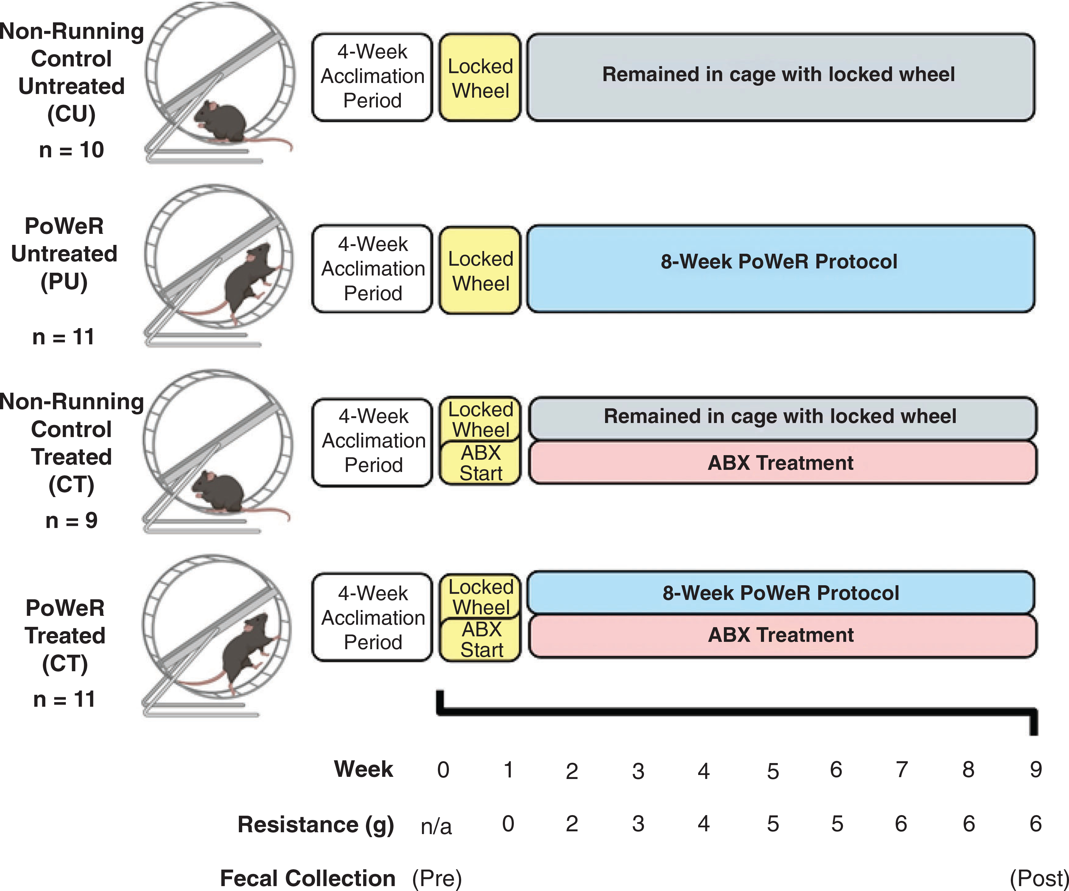
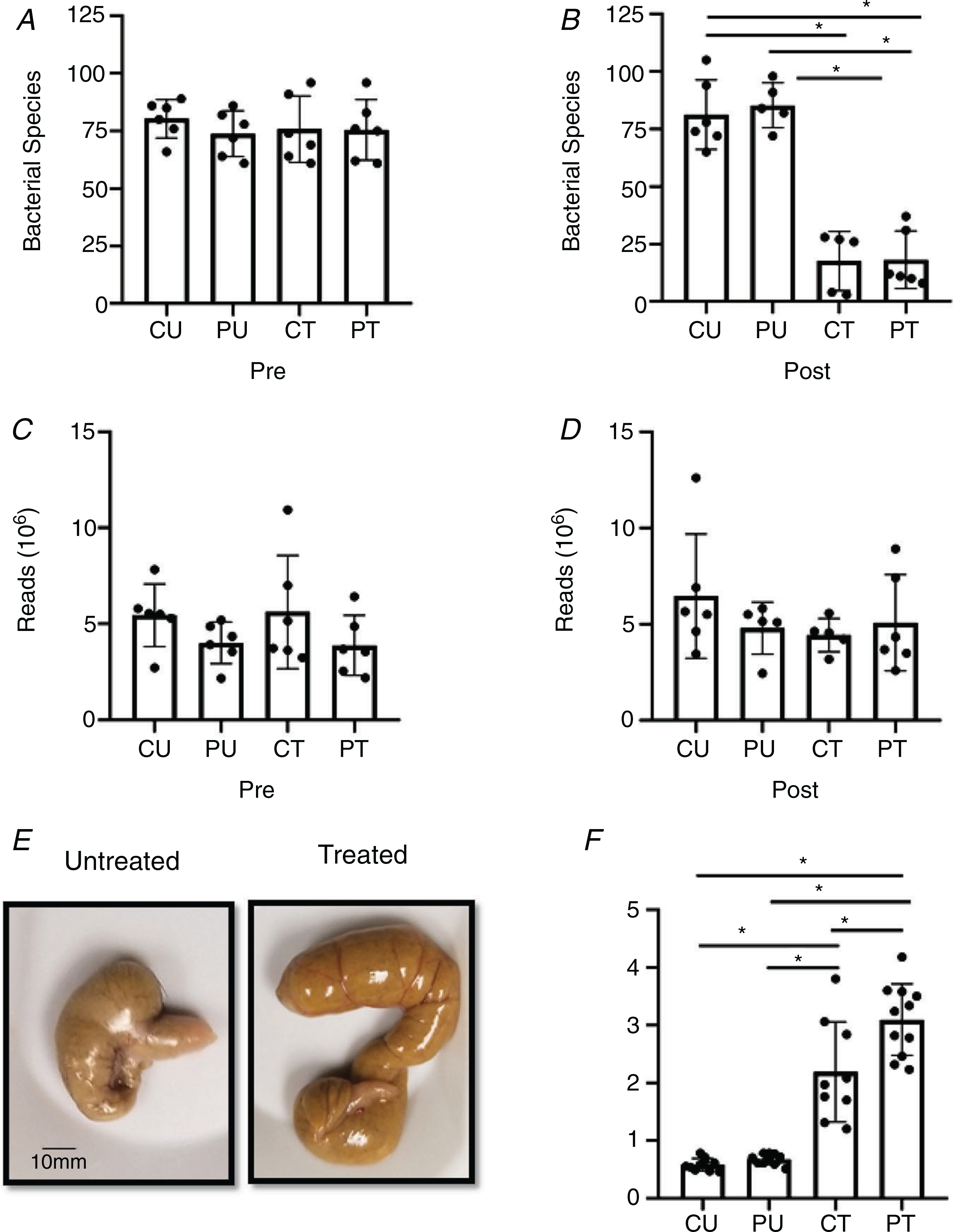
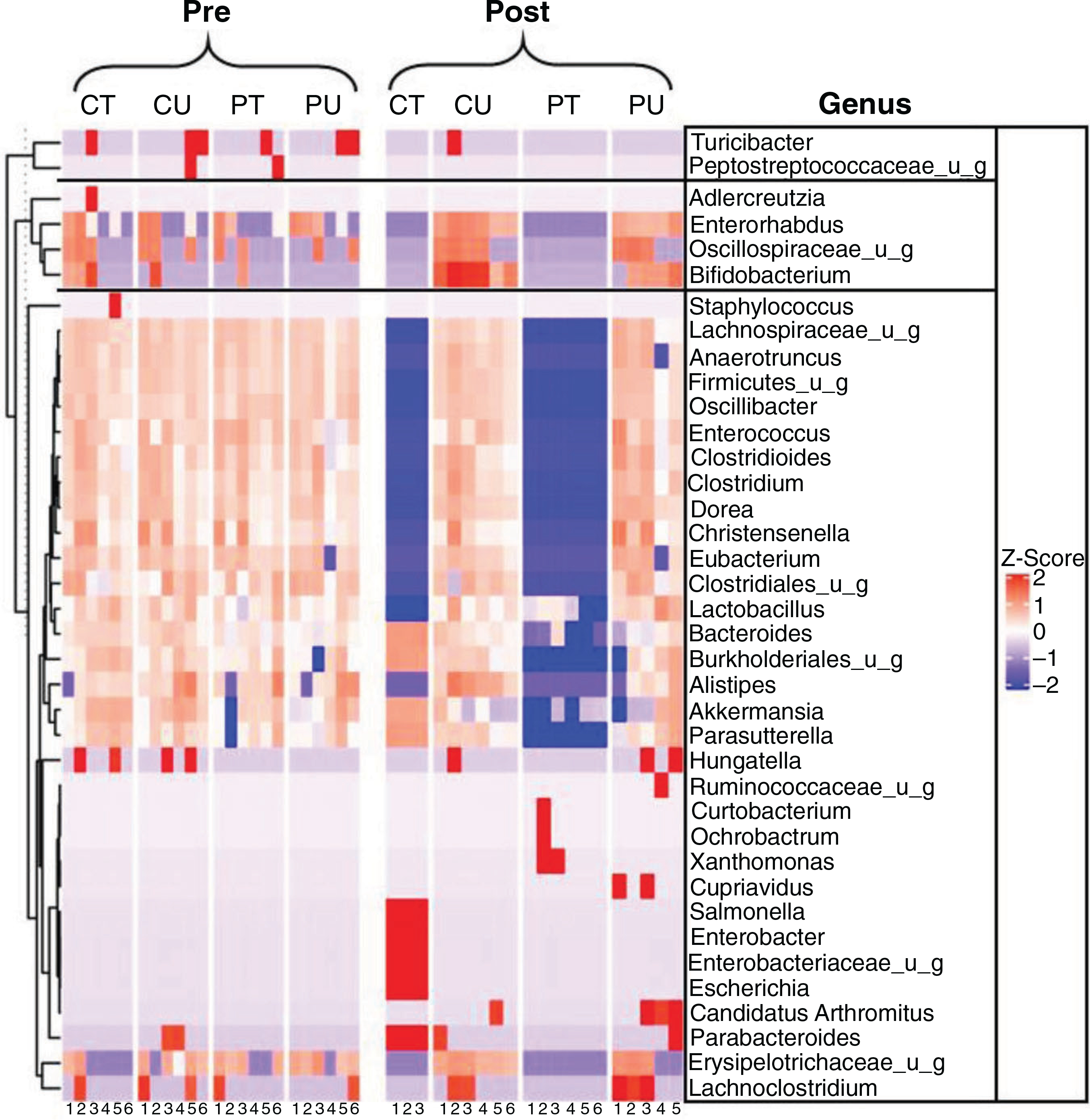
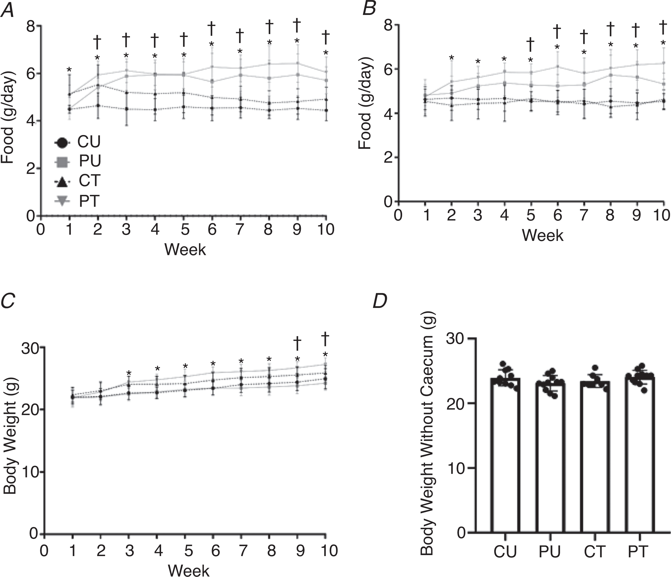
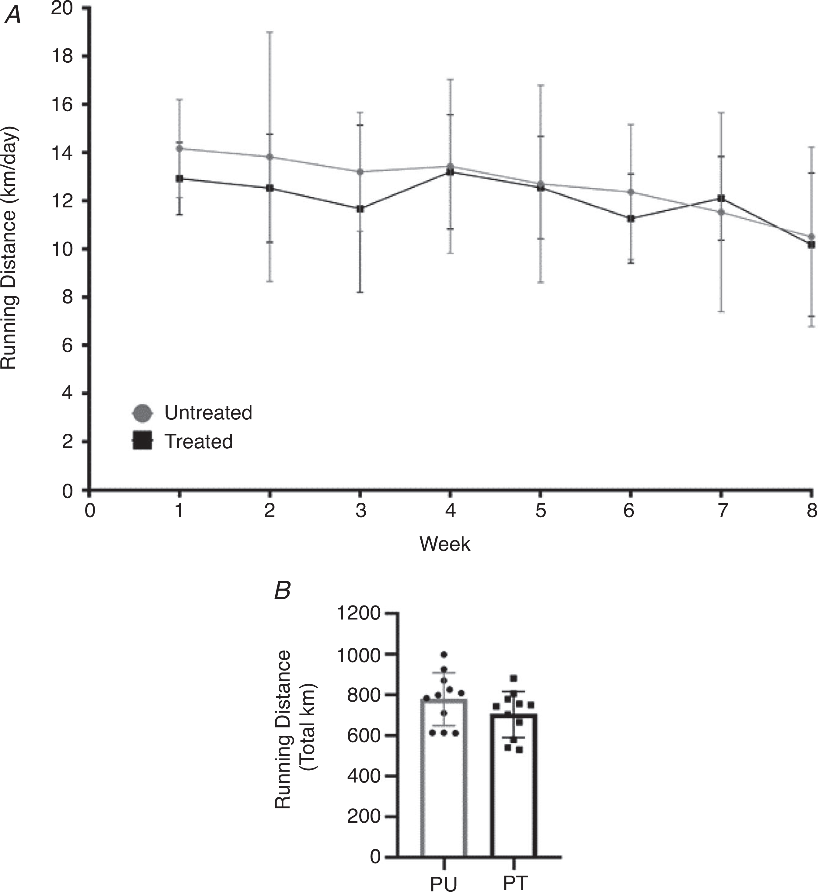
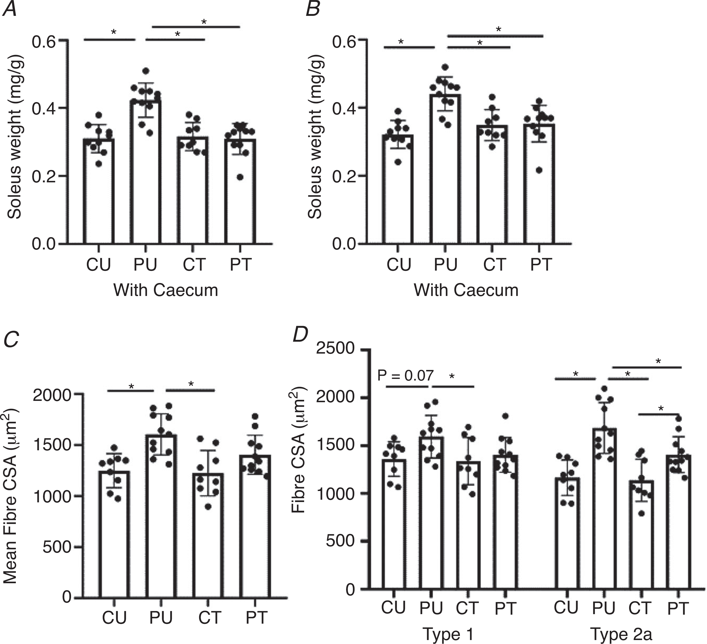
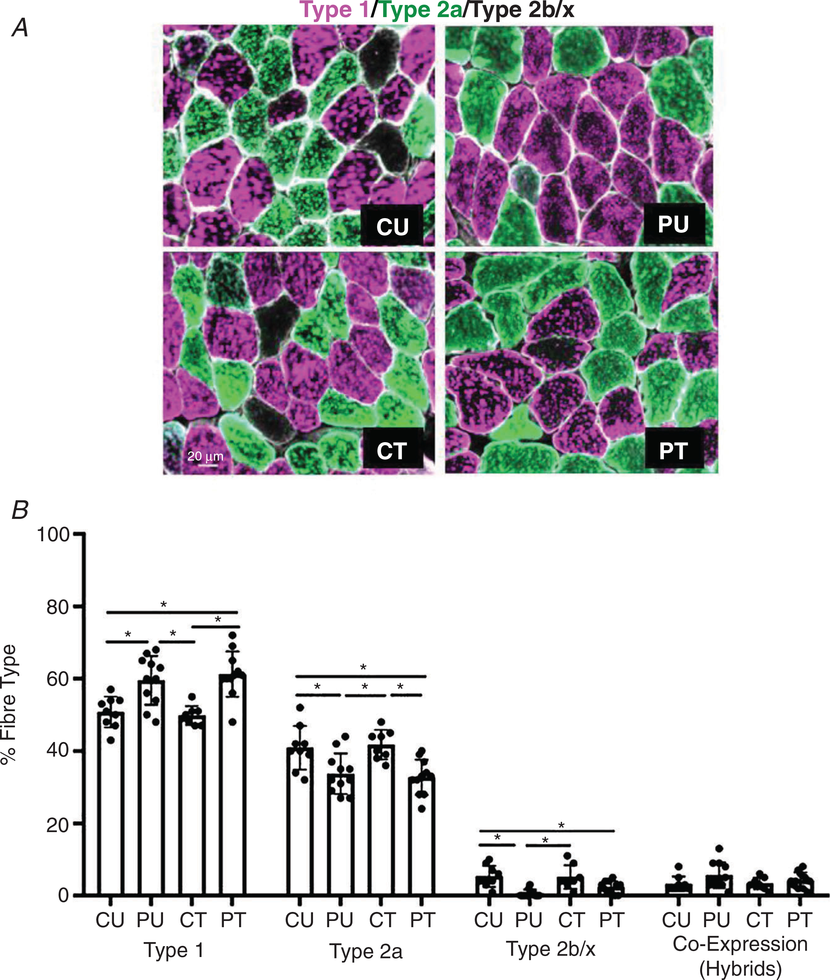
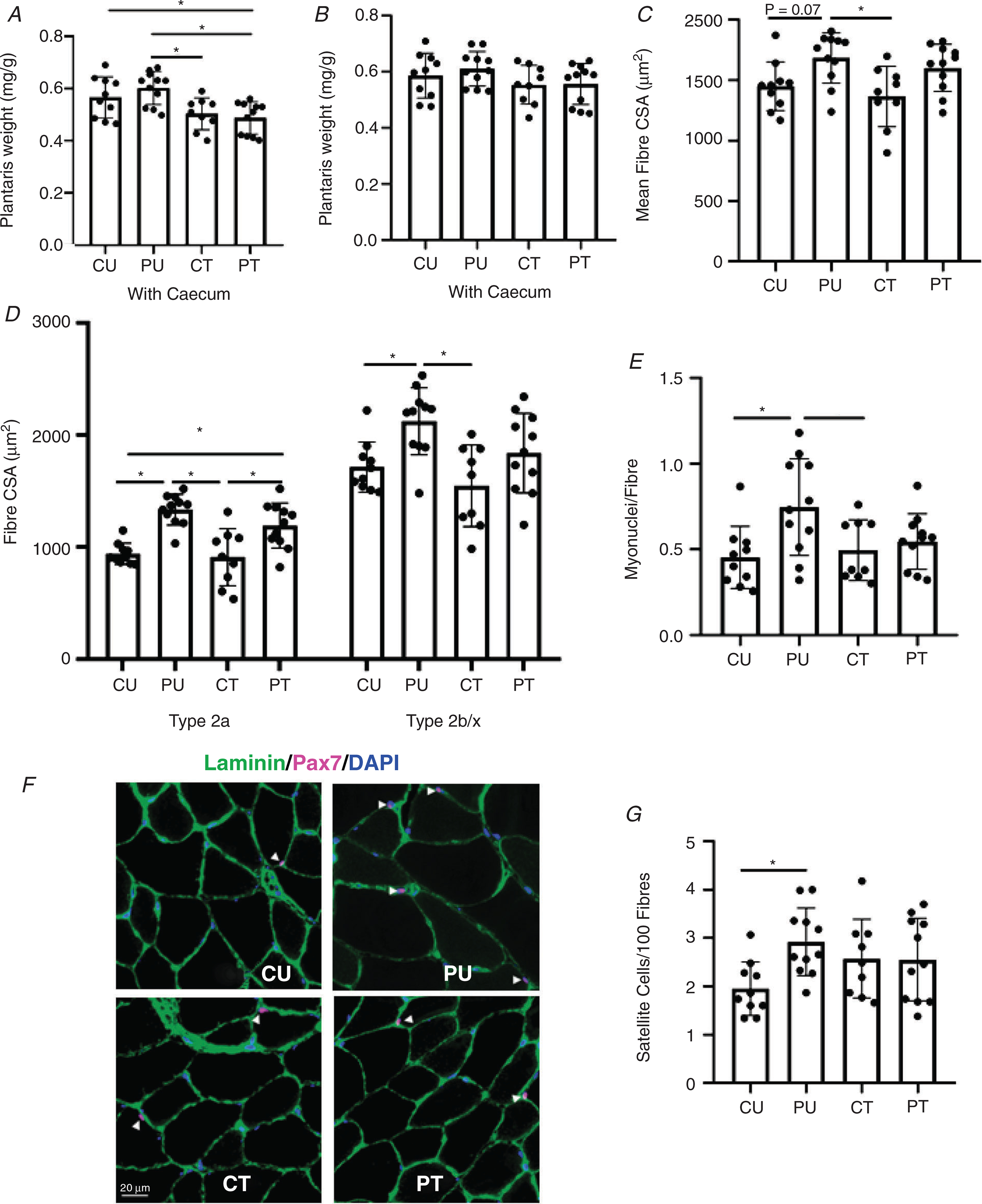
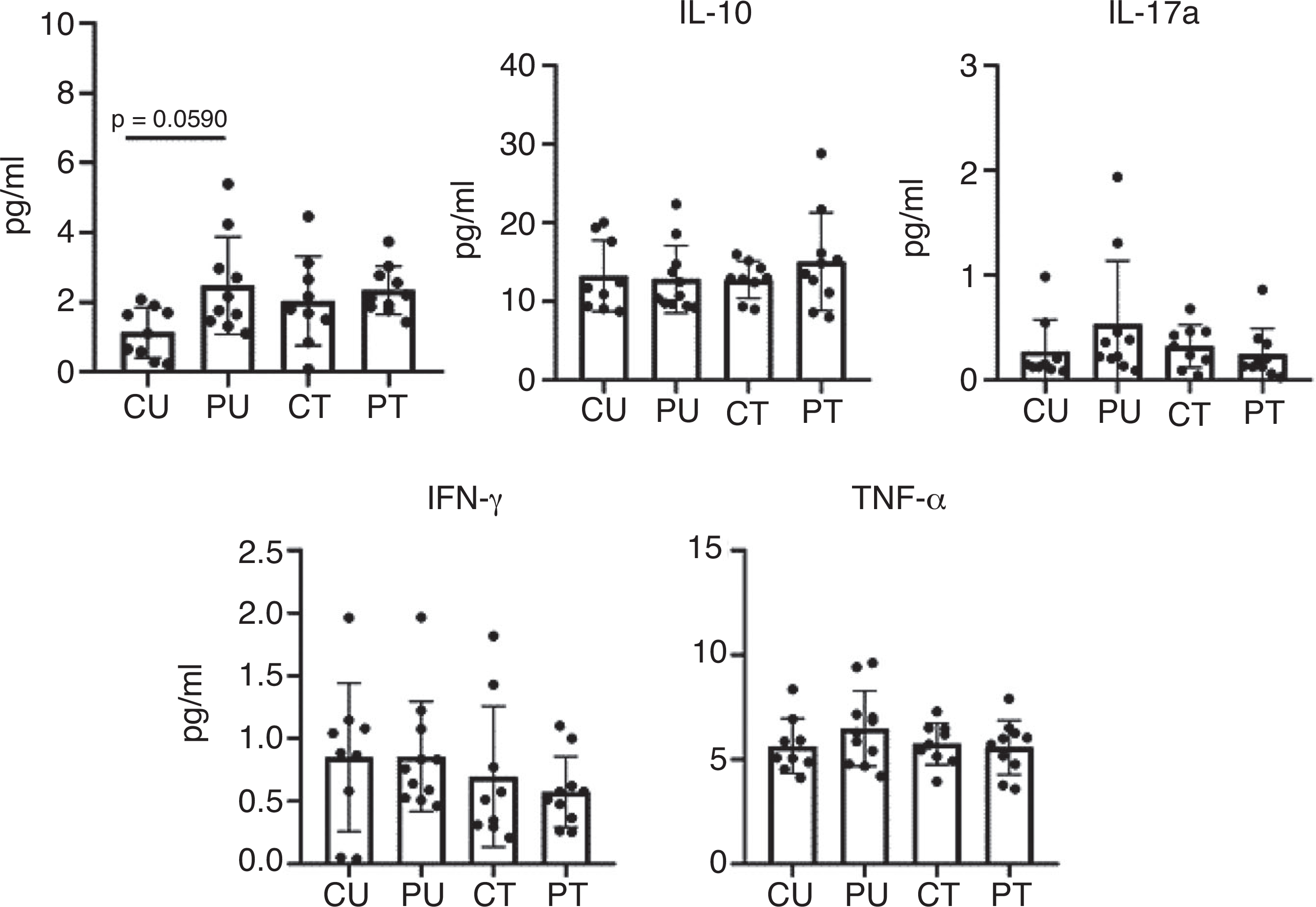
There is emerging evidence of a gut microbiome-skeletal muscle axis. The purpose of this study was to determine if an intact gut microbiome was necessary for skeletal muscle adaptation to exercise. Forty-two 4-month-old female C57BL/6J mice were randomly assigned to untreated (U) or antibiotic-treated (T) non-running controls (CU or CT, respectively) or progressive weighted wheel running (PoWeR, P) untreated (PU) or antibiotic-treated (PT) groups. Antibiotic treatment resulted in disruption of the gut microbiome as indicated by a significant depletion of gut microbiome bacterial species in both CT and PT groups. The training stimulus was the same between PU and PT groups as assessed by weekly (12.35 ± 2.06 vs. 11.09 ± 1.76 km/week, respectively) and total (778.9 ± 130.5 vs. 703.8 ± 112.9 km, respectively) running activity. In response to PoWeR, PT showed less hypertrophy of soleus type 1 and 2a fibres and plantaris type 2b/x fibres compared to PU. The higher satellite cell and myonuclei abundance of PU plantaris muscle after PoWeR was not observed in PT. The fibre-type shift of PU plantaris muscle to a more oxidative type 2a fibre composition following PoWeR was blunted in PT. There was no difference in serum cytokine levels among all groups suggesting disruption of the gut microbiome did not induce systemic inflammation. The results of this study provide the first evidence that an intact gut microbiome is necessary for skeletal muscle adaptation to exercise. KEY POINTS: Dysbiosis of the gut microbiome caused by continuous antibiotic treatment did not affect running activity. Continuous treatment with antibiotics did not result in systemic inflammation as indicated by serum cytokine levels. Gut microbiome dysbiosis was associated with blunted fibre type-specific hypertrophy in the soleus and plantaris muscles in response to progressive weighted wheel running (PoWeR). Gut microbiome dysbiosis was associated with impaired PoWeR-induced fibre-type shift in the plantaris muscle. Gut microbiome dysbiosis was associated with a loss of PoWeR-induced myonuclei accretion in the plantaris muscle.

Keywords: dysbiosis; exercise; gut microbiome; hypertrophy; skeletal muscle.


Conflict of interest statement

Competing interests

All authors declare no competing interests.

Figures

Figure 1.
Figure 1.. Study design
Animals were co-housed in groups of four to five for 4 weeks in order to allow for acclimation to the animal facility at the University of Kentucky. Upon the completion of the acclimation period, mice were randomly split into four different groups and singly housed in running wheel cages. Immediately after randomization into study groups, the first faecal samples were collected (pre), prior to antibiotic administration. During the first week of being singly housed, all wheels were locked, and the antibiotic treatment began. One week after, the wheels were unlocked for those mice in the PoWeR groups, initiating the acclimation week. After the first week of acclimation with an unloaded wheel, 2 g was placed on one side of the running wheel to add resistance Each week thereafter, an additional 1 g was added to the wheel until the load reached a total of 6 g for the final 3 weeks of training. A final faecal sample was collected roughly 48 h prior to euthanasia. After completion of 8 weeks of PoWeR training, mice were euthanized, and tissues were collected for analysis. Image created with BioRender.
Figure 2.
Figure 2.. Gut microbial dysbiosis with antibiotics
Antibiotic induced dysbiosis of the gut microbiome. A and B, number of individual bacterial species before (pre) (A) and after (post) (B) antibiotic treatment and PoWeR training n = 5–6 per group. C and D, number of reads during sequencing corresponding to the pre (C) and post (D) time points; n = 5–6 per group. E, representative image of a caecum harvested from an untreated and treated mouse. F, differences in caeca weight between the groups; n = 9–11 per group. Bars are mean values; circles represent individual values. Errors bars show standard deviation. *P < 0.05.
Figure 3.
Figure 3.. PoWeR- and antibiotic-induced changes to the microbiome composition
Heat map indicating the microbial composition at the genus levels between groups at the pre and post time points. n = 3–6 per group.
Figure 4.
Figure 4.. PoWeR training, not dysbiosis of the gut microbiome, leads to increased food and water consumption but not body weight changes
Food consumption, water intake and body weight changes during PoWeR training. Weekly food consumption during training (A), weekly water intake during study (B), weekly body weight during study (C), and body weight at the point of sacrifice with the caecum removed (D). Bars are means and circles represent individual values. Points in A–C represent group averages for that week, errors bars show standard deviation. n = 9–11 per group. * Significantly different (P < 0.05) PT vs. CT and †significantly (P < 0.05) different PU vs. CU for food and water intake. *Significantly (P < 0.01) different PT vs. CU and PU and †significantly (P < 0.05) different CT vs. PU for body weights.
Figure 5.
Figure 5.. Antibiotic induced dysbiosis of the gut microbiome does not impair exercise activity
Running volume during PoWeR training. A, weekly running volume (km/day) of the PoWeR untreated and treated groups. B, mean total distance run during PoWeR training between PoWeR untreated and treated groups. n = 11 per group. Bars represent means, circles represent individual mice, filled circles in A represent group averages for the corresponding week and error bars show standard deviation.
Figure 6.
Figure 6.. Antibiotic-induced dysbiosis of the gut microbiome results in a blunted hypertrophic response in the soleus
Analysis of the soleus muscle after PoWeR training with or without dysbiosis. A, normalized soleus wet weight to body weight including the caecum. B, normalized soleus wet weight to body weight excluding the caecum. C, soleus mean cross-sectional area (7 μm sections). D, type 1 and type 2a skeletal muscle fibre cross-sectional area (7 μm sections). n = 8–11 per group. Bars represent means, circles represent individual mice and error bars show standard deviation. *P < 0.05.
Figure 7.
Figure 7.. Antibiotic-induced dysbiosis of the gut microbiome does not impair skeletal muscle fibre-type shift in response to PoWeR training in the soleus
Analysis of the fibre-type distribution in the soleus muscle after PoWeR training. A, representative image of soleus cross section. B, fibre-type distribution for type 1, type 2a, type 2b/x and co-expression (hybrid) skeletal muscle fibres. n = 7–11 per group. Bars represent means, circles represent individual mice and error bars show standard deviation. *P < 0.05.
Figure 8.
Figure 8.. Antibiotic-induced dysbiosis of the gut microbiome results in blunted hypertrophy, myonuclei accretion and altered satellite cell abundance in the plantaris muscle
Analysis of the plantaris muscle after PoWeR training. A, normalized plantaris wet weight to body weight including the caecum. B, normalized plantaris wet weight to body weight excluding the caecum. C, plantaris mean fibre cross-sectional area (7 μm sections). D, type 2a and type 2b/x skeletal muscle fibre cross-sectional area (7 μm sections). E, number of myonuclei per fibre. F, representative image of Pax 7+ myonuclei. G, satellite cells per 100 muscle fibres. n = 9–11 per group. Bars represent means, circles represent individual mice and error bars show standard deviation. *P < 0.05.
Figure 9.
Figure 9.. Antibiotic-induced dysbiosis of the gut microbiome results in a blunted fibre-type shift in the plantaris
Analysis of the fibre-type distribution in the plantaris muscle after PoWeR training. A, representative image of plantaris cross section. B, fibre-type distribution for type 1, type 2a, type 2b/x and co-expression (hybrid) skeletal muscle fibres. n = 9–11 per group. Bars represent means, circles represent individual mice and error bars show standard deviation. *P < 0.05.
Figure 10.
Figure 10.. Ten weeks of antibiotic administration and PoWeR training did not augment inflammation
Serum concentrations of inflammatory markers taken after 9 weeks of training and antibiotic administration. n = 9–11 per group. Bars represent means, circles represent individual mice and error bars show standard deviation. IL-6, IL-10 and TNF-α were log transformed.

Comment in

References

    1. Allen JM, Berg Miller ME, Pence BD, Whitlock K, Nehra V, Gaskins HR, White BA, Fryer JD & Woods JA (2015). Voluntary and forced exercise differentially alters the gut microbiome in C57BL/6J mice. J Appl Physiol 118, 1059–1066. - PubMed
    1. Allen JM, Mailing LJ, Cohrs J, Salmonson C, Fryer JD, Nehra V, Hale VL, Kashyap P, White BA & Woods JA (2018). Exercise training-induced modification of the gut microbiota persists after microbiota colonization and attenuates the response to chemically-induced colitis in gnotobiotic mice. Gut Microbes 9, 115–130. - PMC - PubMed
    1. Backhed F, Manchester JK, Semenkovich CF & Gordon JI (2007). Mechanisms underlying the resistance to diet-induced obesity in germ-free mice. Proc Natl Acad Sci U S A 104, 979–984. - PMC - PubMed
    1. Bastiaanssen TFS, Cussotto S, Claesson MJ, Clarke G, Dinan TG & Cryan JF (2020). Gutted! Unraveling the role of the microbiome in major depressive disorder. Harv Rev Psychiatry 28, 26–39. - PMC - PubMed
    1. Bayer F, Ascher S, Pontarollo G & Reinhardt C (2019). Antibiotic treatment protocols and germ-free mouse models in vascular research. Front Immunol 10, 2174. - PMC - PubMed

Publication types