. 2021 Oct 5;10(19):e021542.
doi: 10.1161/JAHA.121.021542. Epub 2021 Sep 25.

Intrahepatic Dynamic Contrast-Enhanced Magnetic Resonance Lymphangiography: Potential Imaging Signature for Protein-Losing Enteropathy in Congenital Heart Disease

Affiliations

Intrahepatic Dynamic Contrast-Enhanced Magnetic Resonance Lymphangiography: Potential Imaging Signature for Protein-Losing Enteropathy in Congenital Heart Disease

Bethan A Lemley et al. J Am Heart Assoc. .

Abstract

Background Protein-losing enteropathy (PLE) is a significant cause of morbidity and mortality in congenital heart disease patients with single ventricle physiology. Intrahepatic dynamic contrast-enhanced magnetic resonance lymphangiography (IH-DCMRL) is a novel diagnostic technique that may be useful in characterizing pathologic abdominal lymphatic flow in the congenital heart disease population and in diagnosing PLE. The objective of this study was to characterize differences in IH-DCMRL findings in patients with single ventricle congenital heart disease with and without PLE. Methods and Results This was a single-center retrospective study of IH-DCMRL findings and clinical data in 41 consecutive patients, 20 with PLE and 21 without PLE, with single ventricle physiology referred for lymphatic evaluation. There were 3 distinct duodenal imaging patterns by IH-DCMRL: (1) enhancement of the duodenal wall with leakage into the lumen, (2) enhancement of the duodenal wall without leakage into the lumen, and (3) no duodenal involvement. Patients with PLE were more likely to have duodenal involvement on IH-DCMRL than patients without PLE (P<0.001). Conclusions IH-DCMRL findings of lymphatic enhancement of the duodenal wall and leakage of lymph into the duodenal lumen are associated with PLE. IH-DCMRL is a useful new modality for characterizing pathologic abdominal lymphatic flow in PLE and might be useful as a risk-assessment tool for PLE in at-risk patients.

Keywords: magnetic resonance lymphangiography; protein‐losing enteropathy; single ventricle heart defects; total cavopulmonary connection.


Conflict of interest statement

Dr Ravishankar has participated in advisory boards on nutrition for Nutricia. The remaining authors have no disclosures to report.

Figures

Figure 1
Figure 1. Intrahepatic dynamic contrast‐enhanced magnetic resonance lymphangiography coronal sections in single ventricle heart disease patients.
A, Duodenal wall enhancement and leakage of contrast into the bowel lumen (arrow) in a patient with PLE. Contrast distribution subsequently visualized throughout bowel. B, Duodenal wall enhancement without leakage of contrast into the bowel lumen (arrow) in a patient with PLE. C, No duodenal wall enhancement or leakage into the bowel lumen (arrow) in a patient without PLE. PLE indicates protein‐losing enteropathy.
Figure 2
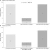
Figure 2. Distributions of IH‐DCMRL imaging patterns (duodenal wall enhancement with leakage into the bowel lumen, duodenal wall enhancement with no leakage into the bowel lumen, and no duodenal involvement) were significantly different between the PLE patients and the no‐PLE patients.
A, Patients at any surgical stage (P<0.001) (B) patients post TCPC (P<0.001). Bars are labeled with counts. IH‐DCMRL indicates intrahepatic dynamic contrast‐enhanced magnetic resonance lymphangiography; PLE, protein‐losing enteropathy; and TCPC, total cavopulmonary connection.
Figure 3
Figure 3. Diagram of the normal hepatic lymphatic networks showing flow from the liver towards the cisterna chyli and into the thoracic duct (A) and hepatic lymphatic flow in PLE showing hepatoduodenal connections to the proximal duodenum (B).
Inset: Dilated lacteals in the duodenal wall are prone to rupture, spilling lymph into the bowel lumen. PLE indicates protein‐losing enteropathy.

References

    1. Goldberg DJ, Dodds K, Rychik J. Rare problems associated with the Fontan circulation. Cardiol Young. 2010;20:113–119. DOI: 10.1017/S1047951110001162. - DOI - PubMed
    1. Allen KY, Downing TE, Glatz AC, Rogers LS, Ravishankar C, Rychik J, Fuller S, Montenegro LM, Steven JM, Spray TL, et al. Effect of Fontan‐associated morbidities on survival with intact Fontan circulation. Am J Cardiol. 2017;119:1866–1871. DOI: 10.1016/j.amjcard.2017.03.004. - DOI - PubMed
    1. Feldt RH, Driscoll DJ, Offord KP, Cha RH, Perrault J, Schaff HV, Puga FJ, Danielson GK. Protein‐losing enteropathy after the Fontan operation. J Thorac Cardiovasc Surg. 1996;112:672–680. DOI: 10.1016/S0022-5223(96)70051-X. - DOI - PubMed
    1. John AS, Johnson JA, Khan M, Driscoll DJ, Warnes CA, Cetta F. Clinical outcomes and improved survival in patients with protein‐losing enteropathy after the Fontan operation. J Am Coll Cardiol. 2014;64:54–62. DOI: 10.1016/j.jacc.2014.04.025. - DOI - PubMed
    1. Mertens L, Hagler DJ, Sauer U, Somerville J, Gewillig M. Protein‐losing enteropathy after the Fontan operation: an international multicenter study. J Thorac Cardiovasc Surg. 1998;115:1063–1073. DOI: 10.1016/S0022-5223(98)70406-4. - DOI - PubMed

Publication types

MeSH terms