. 2021 Dec;12(1):7235-7247.
doi: 10.1080/21655979.2021.1973875.

Role of Fucoxanthin towards Cadmium-induced renal impairment with the antioxidant and anti-lipid peroxide activities

Affiliations

Role of Fucoxanthin towards Cadmium-induced renal impairment with the antioxidant and anti-lipid peroxide activities

Haoyue Yang et al. Bioengineered. 2021 Dec.

Abstract

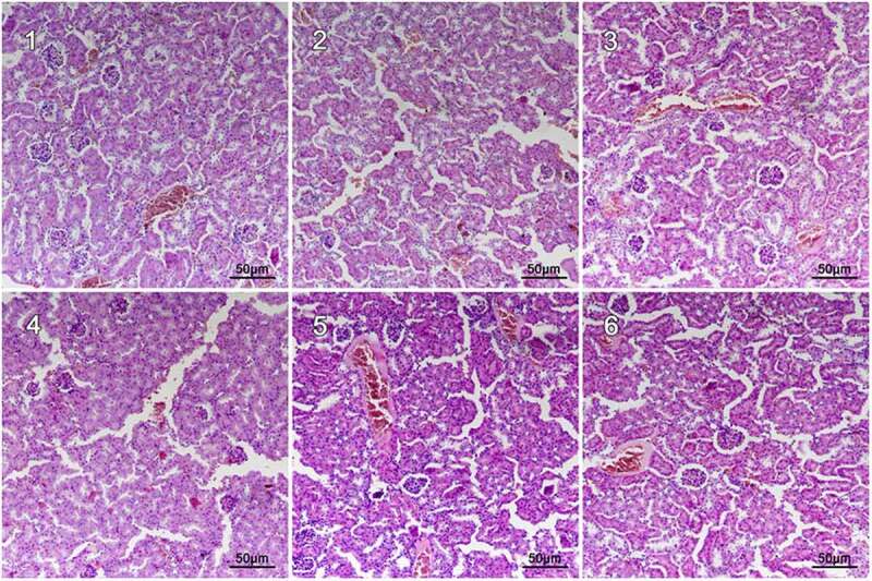
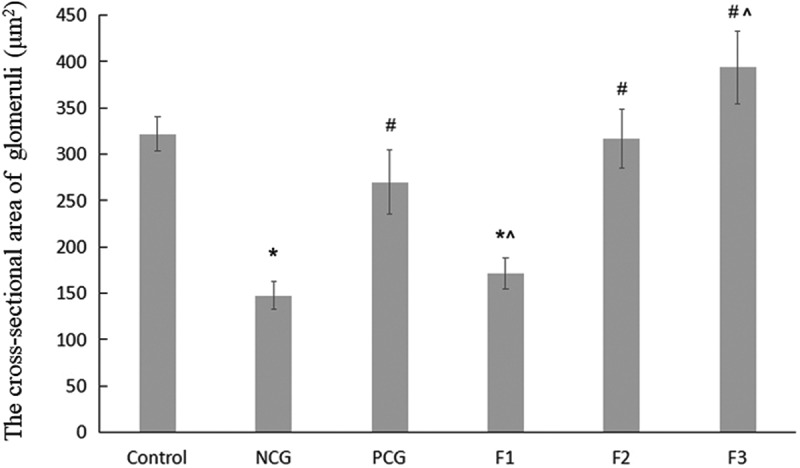
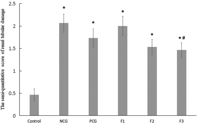
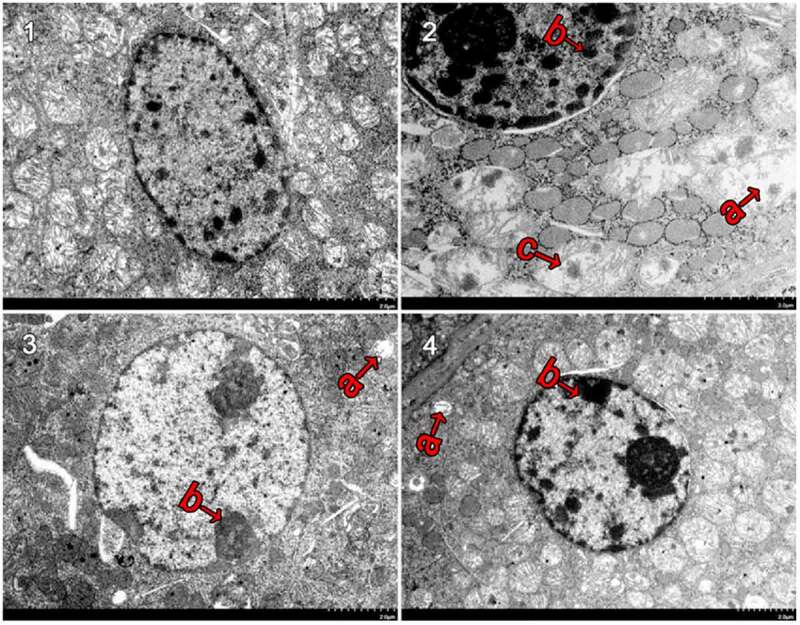
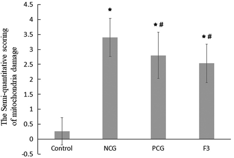
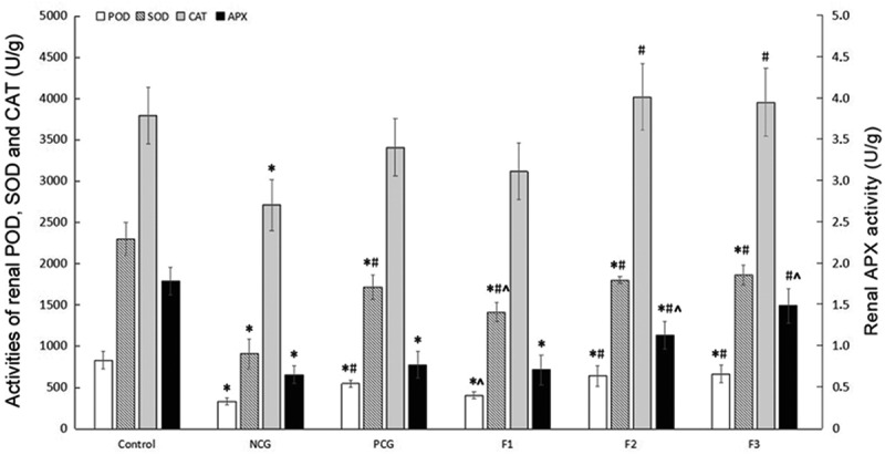
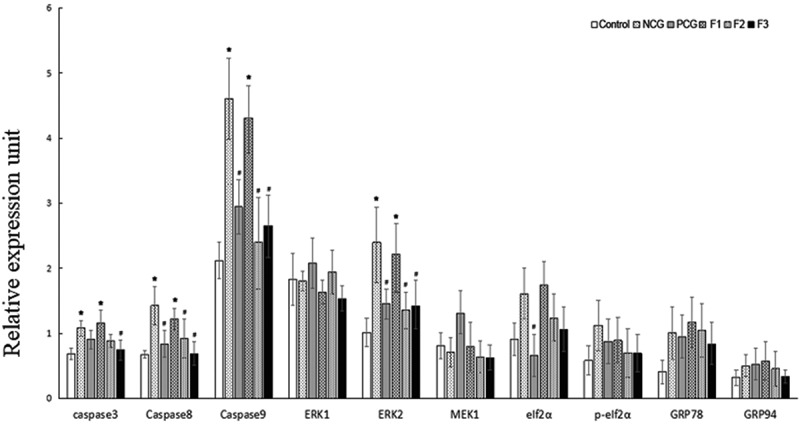
Kidney damages caused by cadmium are considered to be one of the most dangerous consequences for the human body. This study aimed to investigate the protective effects of fucoxanthin supplementation on mice models subjected to cadmium-induced kidney damage. The mice treated with cadmium chloride (CdCl2) were observed to have significantly reduced the cross-section area of glomeruli. Cadmium exposure has also caused the damage of the structural integrity of mitochondria and increased blood urea nitrogen (BUN), kidney injury molecule 1 (KIM1), and neutrophil gelatinase associated lipocalin (NGAL) levels. Peroxidase (POD), superoxide dismutase (SOD), catalase (CAT), and ascorbate peroxidase (APX) levels in cadmium-exposed mice were markedly declined. Caspase3, caspase8, and caspase9 gene expressions in association with apoptosis were dramatically elevated in renal tissues. The CdCl2 treated mice were orally administered with 50 mg/kg Shenfukang, 10 mg/kg, 25 mg/kg, and 50 mg/kg fucoxanthin for 14 days. The results revealed that high doses of fucoxanthin administration significantly decreased BUN, KIM1, NGAL levels, increasing POD, SOD, CAT, and ascorbate APX levels. Fucoxanthin administration also promoted recovery of the renal functions, micro-structural organization, and ultra-structural organization in the renal cells. In summary, the ameliorative effects of fucoxanthin supplementation against cadmium-induced kidney damage were mediated via inhibiting oxidative stress and apoptosis, promoting the recovery of structural integrity of mitochondria.

Keywords: Apoptosis; cadmium chloride; fucoxanthin; oxidative stress; renal function.


Figures

Figure 1.
Figure 1.
Hematoxylin and eosin (H &E) staining of renal in CdCl2 exposure after the treatment with fucoxanthin and Shenfukang tablets supplementation (1) the control group: no CdCl2 administration; (2) NCG, negative control group, only CdCl2 treatment; (3) PCG, positive control group, CdCl2+ Shenfukang tablets; (4) F1, CdCl2 + 10 mg/kg fucoxanthin body weight for 14 day treatment ; (5) F2, CdCl2 + 25 mg/kg body weight fucoxanthin; (6) F3,CdCl2 + 50 mg/kg body weight fucoxanthin. Each group used 20 mice
Figure 2.
Figure 2.
The quantity of the cross-sectional area of the glomeruli (μm2) Single factor analysis of variance between multiple groups was performed using Duncan methods. ‘*’ compared with the control group, P < 0.05; ‘#’ compared with the negative control group, P < 0.05; ‘^’ compared with the positive control group, P < 0.05. Each group used 20 mice
Figure 3.
Figure 3.
The semi-quantitative score of renal tubular damage in mice Single factor analysis of variance between multiple groups was performed using Duncan methods. ‘*’ compared with the control group, P < 0.05; ‘#’ compared with the negative control group, P < 0.05. Each group used 20 mice
Figure 4.
Figure 4.
Ultrastructural changes after treatment with fucoxanthin and Shenfukang tablets administration (1) Control group, (2) NCG, (3) PCG, (4) F3, (a) mitochondria damage, (b) karyopyknosis, and (c) mitochondrial rupture. Each group used 20 mice
Figure 5.
Figure 5.
Semi-quantitative evaluation of mitochondrial damage in mouse kidney cells Single factor analysis of variance between multiple groups was performed using Duncan methods.‘*’ compared with the control group, P < 0.05; ‘#’ compared with the negative control group, P < 0.05. Each group used 20 mice
Figure 6.
Figure 6.
Changes in Renal POD, SOD, CAT and APX Activities (U/g) Single factor analysis of variance between multiple groups was performed using Duncan methods. ‘*’ compared with the control group, P < 0.05; ‘#’ compared with the negative control group, P < 0.05; ‘^’ compared with the positive control group, P < 0.05. Each group used 20 mice
Figure 7.
Figure 7.
Effect of fucoxanthin on the expression of apoptosis-associated genes in the cadmium kidney-damaged mouse model Single factor analysis of variance between multiple groups was performed using Duncan methods.‘*’ compared with the control group, P < 0.05; ‘#’ compared with the negative control group, P < 0.05. Each group used 20 mice

Similar articles

Cited by

References

    1. Donangelo CM, Kerr BT, Queirolo EI, et al. Lead exposure and indices of height and weight in Uruguayan urban school children, considering co-exposure to cadmium and arsenic, sex, iron status and dairy intake. Environ Res. 2021;195:110799. - PMC - PubMed
    1. Nawrot T, Plusquin M, Hogervorst J, et al. Environmental exposure to cadmium and risk of cancer: a prospective population-based study. Lancet Oncol. 2006;7(2):119–126. - PubMed
    1. Satarug S, Garrett SH, Sens MA, et al. Cadmium, environmental exposure, and health outcomes. Environ Health Perspect. 2010;118(2):182–190. - PMC - PubMed
    1. Satarug S, Baker JR, Urbenjapol S, et al. A global perspective on cadmium pollution and toxicity in non-occupationally exposed population. Toxicol Lett. 2003;137(1–2):65–83. - PubMed
    1. Gobe GC, Johnson DW.. Distal tubular epithelial cells of the kidney: potential support for proximal tubular cell survival after renal injury. Int J Biochem Cell Biol. 2007;39(9):1551–1561. - PubMed

Publication types