. 2021 Sep 7:(175):10.3791/61100.
doi: 10.3791/61100.

Quantifying Tissue-Specific Proteostatic Decline in Caenorhabditis elegans

Affiliations

Quantifying Tissue-Specific Proteostatic Decline in Caenorhabditis elegans

Maria I Lazaro-Pena et al. J Vis Exp. .

Abstract

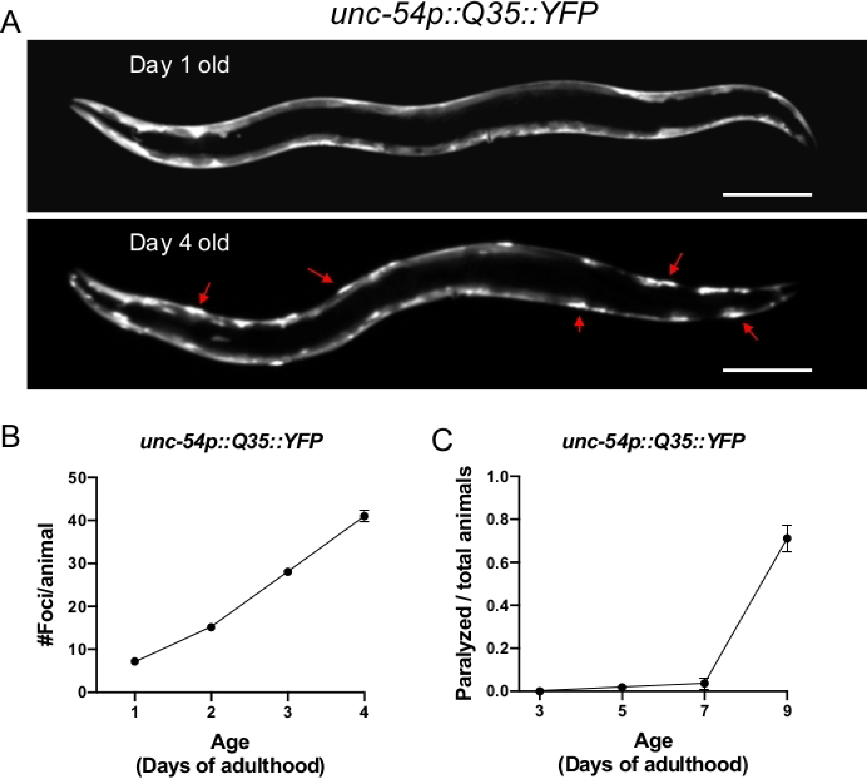
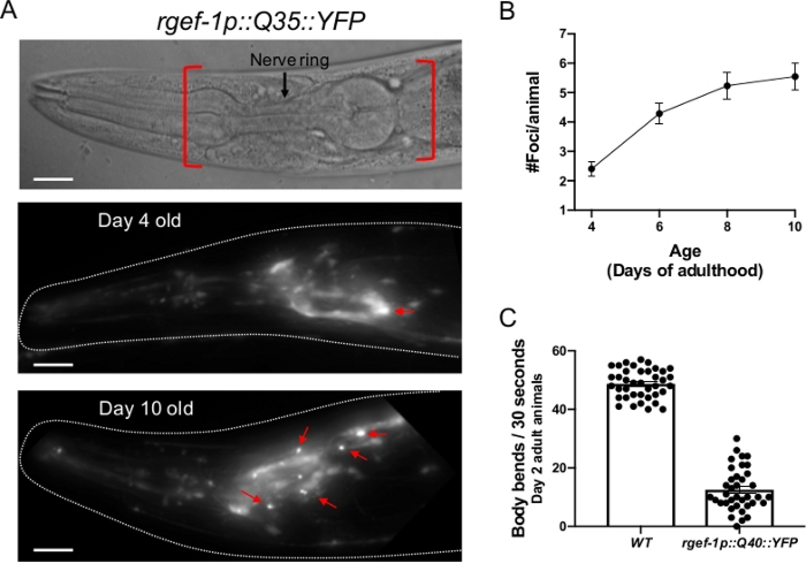
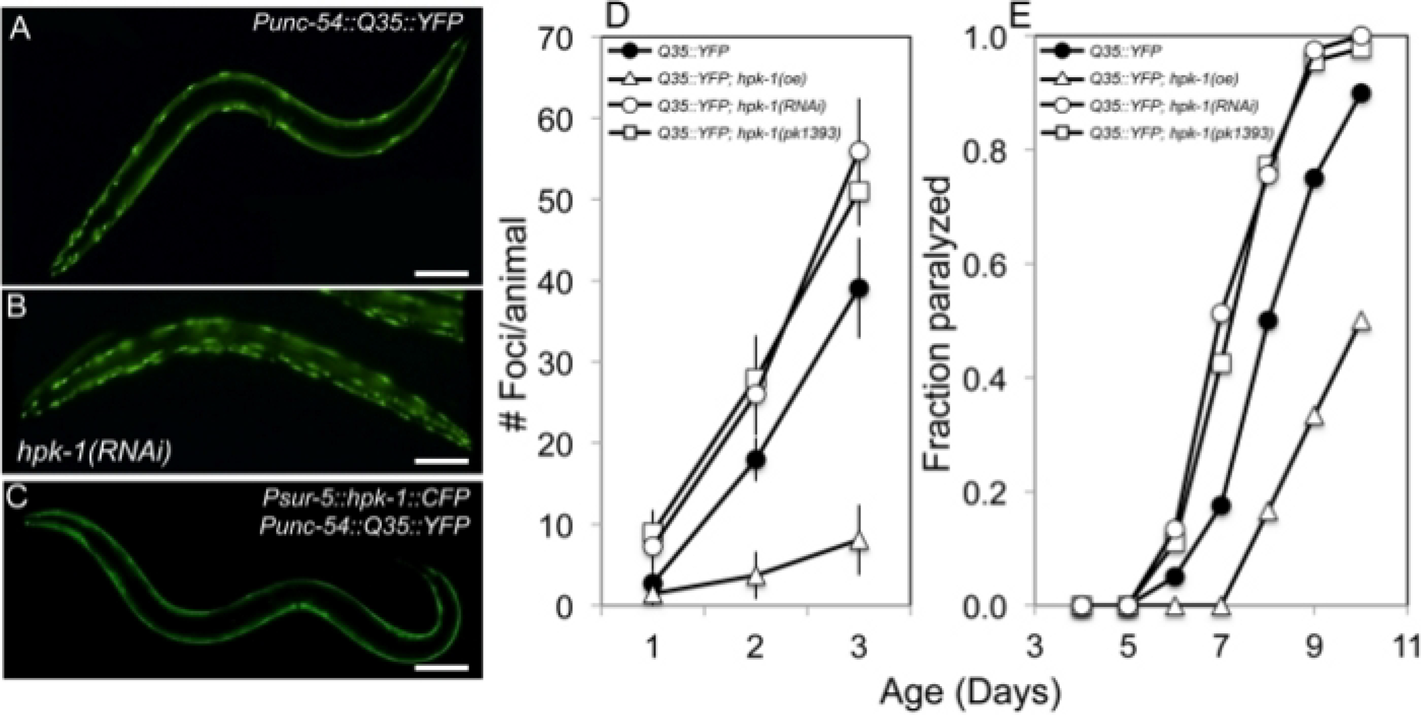
The ability to maintain proper function and folding of the proteome (protein homeostasis) declines during normal aging, facilitating the onset of a growing number of age-associated diseases. For instance, proteins with polyglutamine expansions are prone to aggregation, as exemplified with the huntingtin protein and concomitant onset of Huntington's disease. The age-associated deterioration of the proteome has been widely studied through the use of transgenic Caenorhabditis elegans expressing polyQ repeats fused to a yellow fluorescent protein (YFP). This polyQ::YFP transgenic animal model facilitates the direct quantification of the age-associated decline of the proteome through imaging the progressive formation of fluorescent foci (i.e., protein aggregates) and subsequent onset of locomotion defects that develop as a result of the collapse of the proteome. Further, the expression of the polyQ::YFP transgene can be driven by tissue-specific promoters, allowing the assessment of proteostasis across tissues in the context of an intact multicellular organism. This model is highly amenable to genetic analysis, thus providing an approach to quantify aging that is complementary to lifespan assays. We describe how to accurately measure polyQ::YFP foci formation within either neurons or body wall muscle during aging, and the subsequent onset of behavioral defects. Next, we highlight how these approaches can be adapted for higher throughput, and potential future applications using other emerging strategies for C. elegans genetic analysis.


Conflict of interest statement

Disclosures

The authors declare that they have no competing financial interests.

Figures

Figure 1:
Figure 1:. Expression of polyQ::YFP within C. elegans muscle results in progressive foci accumulation and paralysis during aging.
(A) C. elegans unc-54p::Q35::YFP expression at days 1 and 4 of adulthood (upper and lower panel, respectively). Arrows indicate representative foci. (B) Quantification of fluorescent foci over the first 4 days of adulthood. Foci are resistant to FRAP,,, consistent with an insoluble protein aggregate. Error bars represent standard error of the mean (SEM) (C) unc-54p::Q35::YFP animals become paralyzed during aging. Error bars represent standard error of proportion. Raw data for (B-C) is provided in Supplemental Table 1. The scale bar represents 100 μm in all panels.
Figure 2:
Figure 2:. Expression of polyQ::YFP within C. elegans neurons results in progressive foci accumulation and disruption of normal body bends.
(A, top) DIC image of the C. elegans anterior. The pharynx is a bi-lobed structure in the head of the animal, which is surrounded by the nerve ring, an interconnected cluster of 180 neurons. Red brackets indicate region to score for foci within head neurons. (A, middle) rgefp-1::Q40::YFP fluorescence at day 2 adulthood. Note that YFP expression is largely diffuse, with the exception of an occasional aggregate (arrow). (A, bottom) rgefp-1::Q40::YFP fluorescence at day 10 adulthood. Foci/aggregates are indicated (red arrow). (B) Quantification of fluorescent foci over the first 10 days of adulthood. Foci are resistant to FRAP,,, consistent with an insoluble protein aggregate. Error bars represent standard error of the mean (C) Typical frequency of body bends of wild type and rgef-1p::Q40::YFP animals maintained at 20°C feeding on empty vector RNAi (L4440) at days 2 adulthood. Increased glutamine expansion correlates with movement defects. Error bars represent standard error of the mean. Raw data for (B-C) is provided in Supplemental Table 1. The scale bar represents 20 μm in all panels.
Figure 3:
Figure 3:. HPK-1 promotes proteostasis.
(A-C) hpk-1 activity affects the accumulation of Q35::YFP foci in muscle cells. Shown are representative images of Punc-54::polyQ::YFP animals treated with (A) control RNAi or (B) hpk-1 RNAi, and (C) transgenic animals overexpressing hpk-1 (Psur-5::HPK-1::CFP). (D) Time course of polyQ::YFP foci accumulation in conjunction with: treatment with control RNAi (black circles), hpk-1 RNAi (white circles), hpk-1(pk1393) (white squares), or hpk-1 overexpression (open triangles). Data points display the mean ± standard deviation (S.D.) of at least 15 animals per biological replicate; at least 5 independent experiments were performed. (E) Time course of paralysis of Punc-54::polyQ::YFP animals in conjunction with: treatment with control RNAi (black circles), hpk-1 RNAi (white circles), hpk-1(pk1393) (white squares), or hpk-1 overexpression (open triangles). Plotted data display the results for a single representative trial. This figure is reprinted from reference with permission via a Creative Commons Attribution (CC BY) license. The scale bar represents 100 μm in all panels.

References

    1. Wolff S, Weissman JS, Dillin A Differential scales of protein quality control. Cell. 157 (1), 52–64 (2014). - PubMed
    1. Labbadia J, Morimoto RI The biology of proteostasis in aging and disease. Annual Review of Biochemistry. 84 435–464 (2015). - PMC - PubMed
    1. Powers ET, Morimoto RI, Dillin A, Kelly JW, Balch WE Biological and chemical approaches to diseases of proteostasis deficiency. Annual Review of Biochemistry. 78 959–991 (2009). - PubMed
    1. Brehme M et al. A chaperome subnetwork safeguards proteostasis in aging and neurodegenerative disease. Cell Reports. 9 (3), 1135–1150 (2014). - PMC - PubMed
    1. Sala AJ, Bott LC, Morimoto RI Shaping proteostasis at the cellular, tissue, and organismal level. Journal of Cell Biology. 216 (5), 1231–1241 (2017). - PMC - PubMed

Publication types

LinkOut - more resources