

Measurement of Fatty Acid β-Oxidation in a Suspension of Freshly Isolated Mouse Hepatocytes
- PMID: 34570107
- PMCID: PMC9035282
- DOI: 10.3791/62904
Measurement of Fatty Acid β-Oxidation in a Suspension of Freshly Isolated Mouse Hepatocytes
Abstract
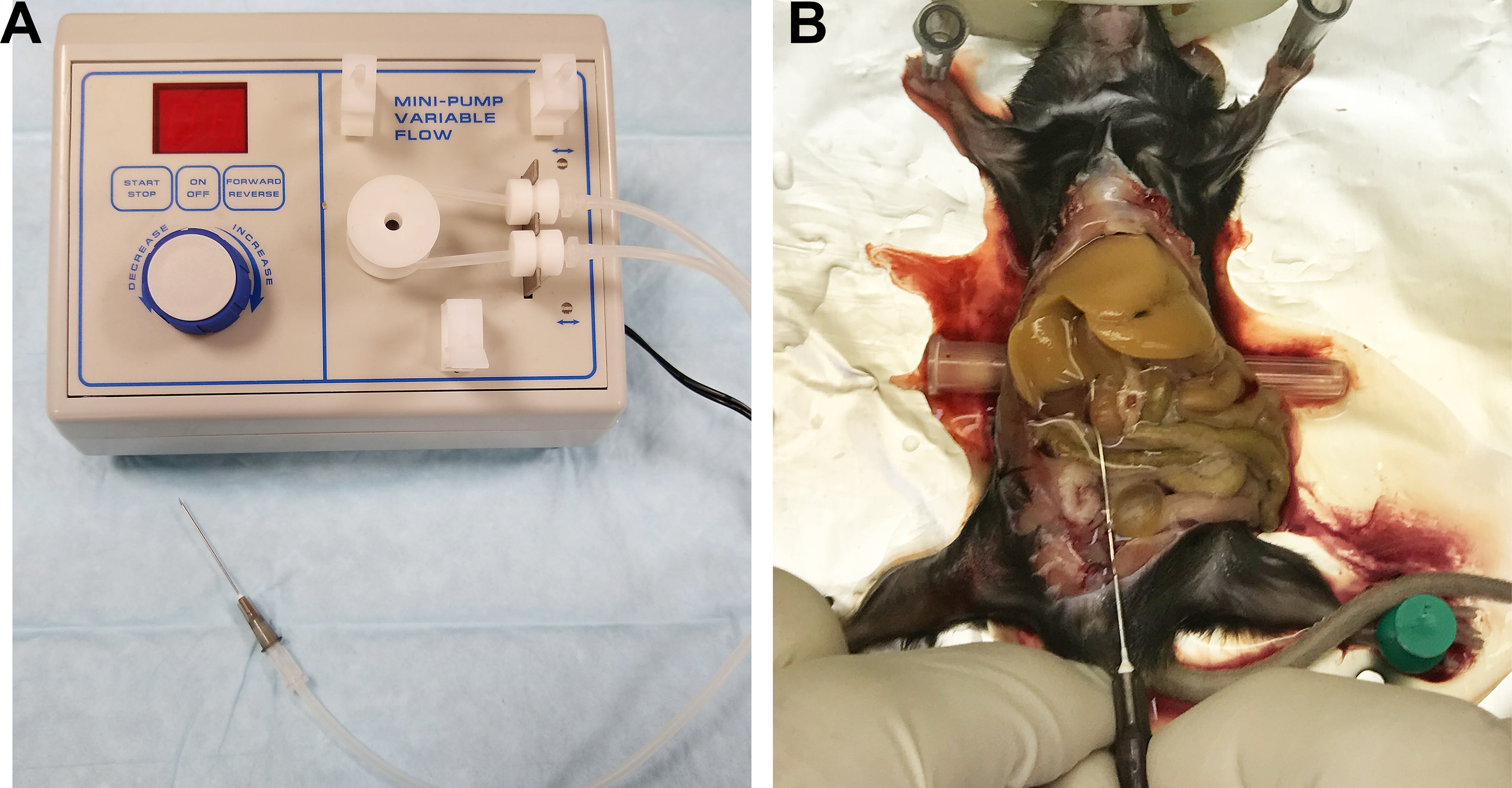
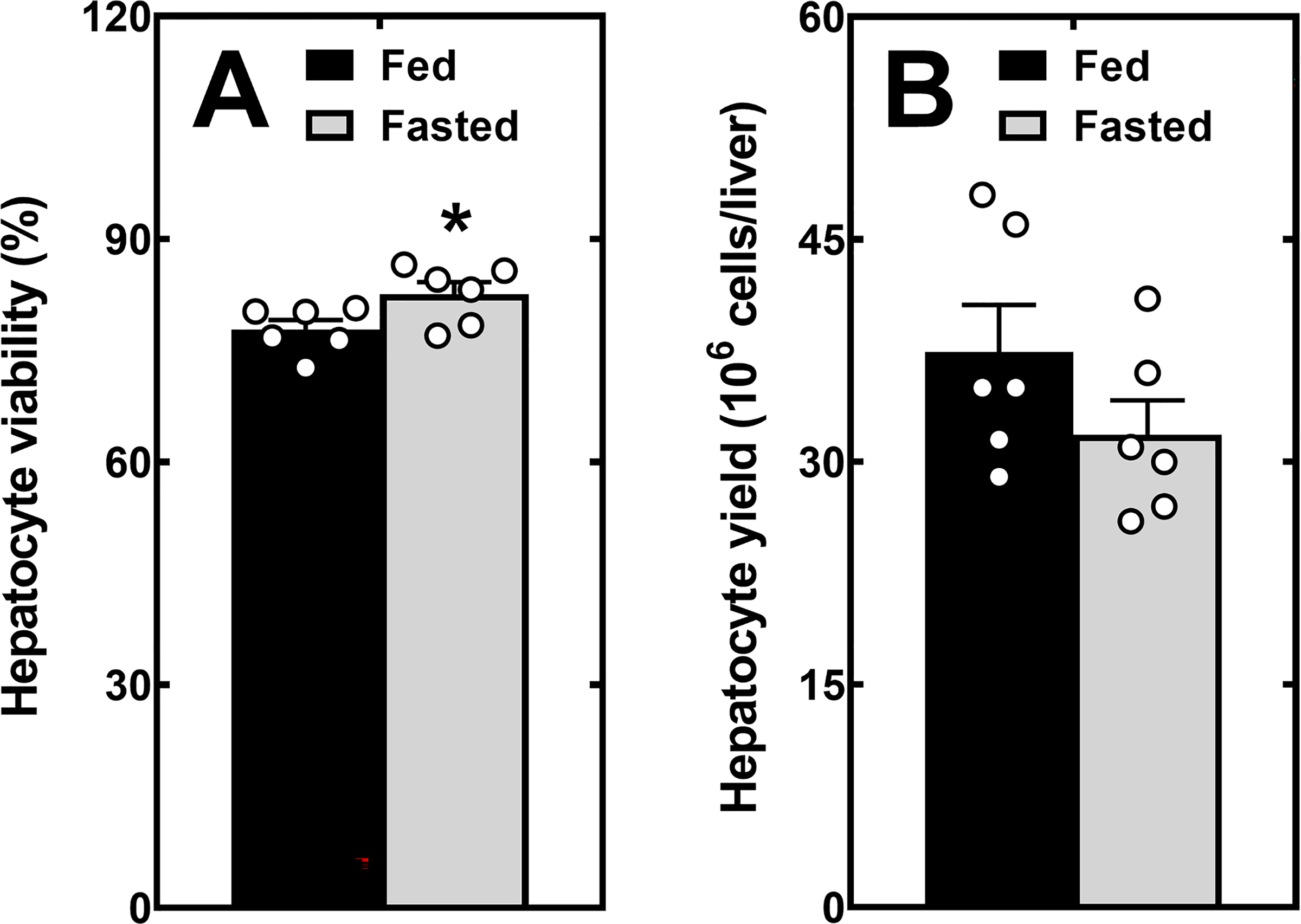
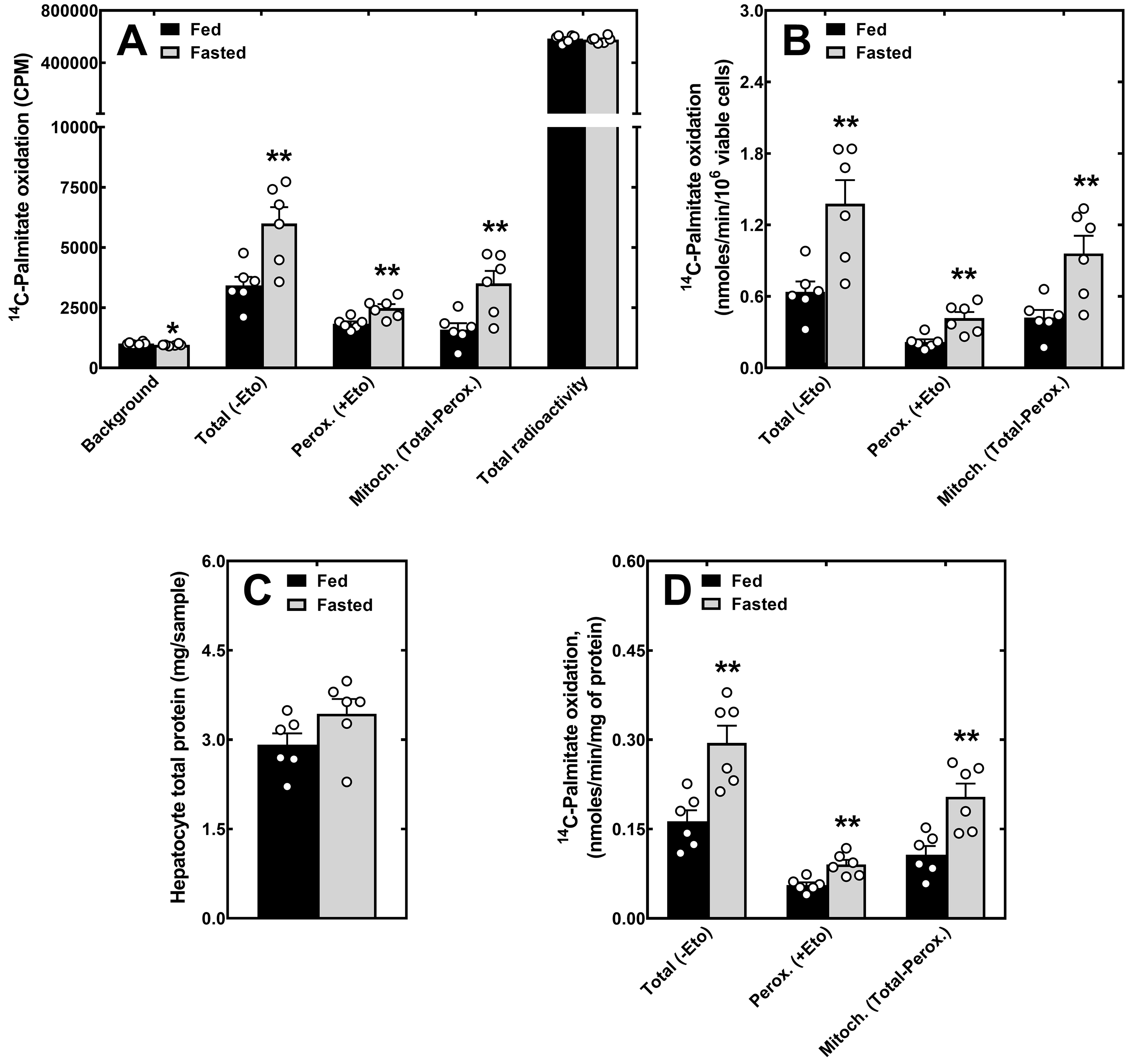
Fatty acid β-oxidation is a key metabolic pathway to meet the energy demands of the liver and provide substrates and cofactors for additional processes, such as ketogenesis and gluconeogenesis, which are essential to maintain whole-body glucose homeostasis and support extra-hepatic organ function in the fasted state. Fatty acid β-oxidation occurs within the mitochondria and peroxisomes and is regulated through multiple mechanisms, including the uptake and activation of fatty acids, enzyme expression levels, and availability of cofactors such as coenzyme A and NAD+. In assays that measure fatty acid β-oxidation in liver homogenates, cell lysis and the common addition of supraphysiological levels of cofactors mask the effects of these regulatory mechanisms. Furthermore, the integrity of the organelles in the homogenates is hard to control and can vary significantly between preparations. The measurement of fatty acid β-oxidation in intact primary hepatocytes overcomes the above pitfalls. This protocol describes a method for the measurement of fatty acid β-oxidation in a suspension of freshly isolated primary mouse hepatocytes incubated with 14C-labeled palmitic acid. By avoiding hours to days of culture, this method has the advantage of better preserving the protein expression levels and metabolic pathway activity of the original liver, including the activation of fatty acid β-oxidation observed in hepatocytes isolated from fasted mice compared to fed mice.
Conflict of interest statement
DISCLOSURES
The authors have no conflicts of interest to disclose.
Figures
Similar articles
-
The role of chicken ovalbumin upstream promoter transcription factor II in the regulation of hepatic fatty acid oxidation and gluconeogenesis in newborn mice.Am J Physiol Endocrinol Metab. 2015 May 15;308(10):E868-78. doi: 10.1152/ajpendo.00433.2014. Epub 2015 Mar 17. Am J Physiol Endocrinol Metab. 2015. PMID: 25783893
-
Effects of benfluorex on fatty acid and glucose metabolism in isolated rat hepatocytes: from metabolic fluxes to gene expression.Diabetes. 2002 Aug;51(8):2363-8. doi: 10.2337/diabetes.51.8.2363. Diabetes. 2002. PMID: 12145146
-
Liver fatty acid binding protein is required for high rates of hepatic fatty acid oxidation but not for the action of PPARalpha in fasting mice.FASEB J. 2004 Feb;18(2):347-9. doi: 10.1096/fj.03-0330fje. Epub 2003 Dec 4. FASEB J. 2004. PMID: 14656998
-
Energy metabolism in the liver.Compr Physiol. 2014 Jan;4(1):177-97. doi: 10.1002/cphy.c130024. Compr Physiol. 2014. PMID: 24692138 Free PMC article. Review.
-
Metabolic adaptations to change of nutrition at birth.Biol Neonate. 1990;58 Suppl 1:3-15. doi: 10.1159/000243294. Biol Neonate. 1990. PMID: 2265217 Review.
Cited by
-
Gemfibrozil-Induced Intracellular Triglyceride Increase in SH-SY5Y, HEK and Calu-3 Cells.Int J Mol Sci. 2023 Feb 3;24(3):2972. doi: 10.3390/ijms24032972. Int J Mol Sci. 2023. PMID: 36769295 Free PMC article.
References
-
- Lopaschuk GD, Ussher JR, Folmes CD, Jaswal JS, Stanley WC Myocardial fatty acid metabolism in health and disease. Physiological Reviews. 90 (1), 207–258, (2010). - PubMed
-
- Mannaerts GP, van Veldhoven PP Functions and organization of peroxisomal beta-oxidation. Annals of the New York Academy of Sciences. 804 99–115, (1996). - PubMed
-
- Kerner J, Hoppel C Fatty acid import into mitochondria. Biochimica et Biophysica Acta (BBA) - Bioenergetics. 1486 (1), 1–17, (2000). - PubMed
Publication types
MeSH terms
Substances
Grants and funding
LinkOut - more resources
Full Text Sources
