

doi: 10.1093/nar/gkab852.
The G protein database, GproteinDb
Affiliations
- PMID: 34570219
- PMCID: PMC8728128
- DOI: 10.1093/nar/gkab852
Item in Clipboard
The G protein database, GproteinDb
Nucleic Acids Res.
.
Abstract
Two-thirds of signaling substances, several sensory stimuli and over one-third of drugs act via receptors coupling to G proteins. Here, we present an online platform for G protein research with reference data and tools for analysis, visualization and design of scientific studies across disciplines and areas. This platform may help translate new pharmacological, structural and genomic data into insights on G protein signaling vital for human physiology and medicine. The G protein database is accessible at https://gproteindb.org.
© The Author(s) 2021. Published by Oxford University Press on behalf of Nucleic Acids Research.
Figures
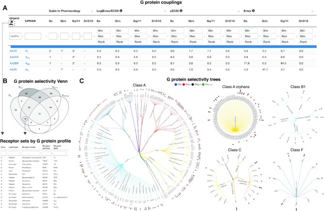
G protein couplings and selectivity. (A) G protein couplings encompass Guide to Pharmacology (GtP) primary/secondary transducers (22) and log(Emax/EC50), pEC50 and Emax values from profiling studies (3–5) and a means thereof. The confidence and coverage of quantitative couplings can be adjusted based on counts of supporting datasets (default 2) and standard deviations from basal signal (default 1.4). (B) G protein selectivity profiles can be intersected in a Venn diagram counting the receptors in each class. (C), G protein selectivity trees map couplings to trees classifying receptors by class, ligand type and receptor families sharing endogenous ligand(s). (B, C) Dashed arrows illustrate paths to obtain coupling-based selection of receptor sets for further study.
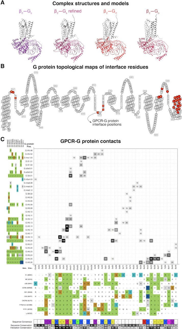
Structures and interface interactions. (A) β2-adrenoceptor-Gs structure (35), its refined structure and β2-Gi1 and -Go models. (B) Snakeplot mapping residues topological segments and highlighting GPCR–G protein interface positions (red residues). (C) Interface interactions shown in % and grayscale across class A GPCR–Gi/o family structures (interaction presence/absences in each structure is shown upon mouse hover). GPCR and G protein residues are indexed with generic residue numbers (25,26) to compare of structurally corresponding positions and color-coded to visualize properties or interaction types. A structure-based interface fingerprint can be profiled against GPCR lacking structures (Supplementary Figure S2).
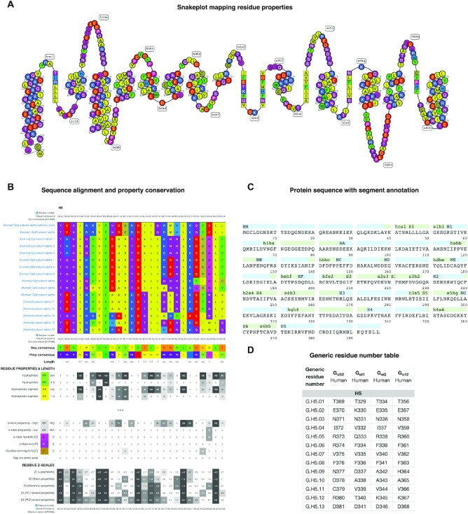
Sequence data in the online G protein research platform. (A) Snakeplot visualization of amino acid topological positions and properties. All amino acids are colored by property: polarity for most but backbone modifying ability for proline and glycine and disulphide-formation for cysteine. (B) Sequence alignment of the receptor-interacting helix 5 of all human 16 Gα proteins along with conservation measures for amino acids and residue groups with similar property and size, and numeric amino acid descriptors, ‘z-scales’ (29) (from https://gproteindb.org/alignment/gproteinselection ). (C) Protein sequence mapping onto segments by secondary structures (helices, β-sheet and loops). (D) Protein-specific and common residue numbers (25) for Gαs, Gαi1, Gαq and Gα12 (from https://gproteindb.org/residue/residuetable_gprot ). These number tables can also be downloaded in Excel format or retrieved programmatically via a RESTFUL-API web service to integrate the numbering in any dataset and analysis method. (A, B) Taken from the ‘G protein page’ (https://gproteindb.org/signprot ) for Gαs. (A–C), Common residue numbers (25) can be shown by mouse hover.
References
-
- Avet C., Mancini A., Breton B., Le Gouill C., Hauser A.S., Normand C., Kobayashi H., Gross F., Hogue M., Lukasheva V.et al. .. Effector membrane translocation biosensors reveal G protein and B-arrestin profiles of 100 therapeutically relevant GPCRs. 2020; bioRxiv doi:24 April 2020, preprint: not peer reviewed10.1101/2020.04.20.052027. - DOI - PMC - PubMed
Publication types
MeSH terms
Substances
LinkOut - more resources
Full Text Sources
Other Literature Sources
