. 2022 Mar;64(3):553-563.
doi: 10.1007/s00234-021-02814-8. Epub 2021 Sep 27.

Hemodynamic evaluation of patients with Moyamoya Angiopathy: comparison of resting-state fMRI to breath-hold fMRI and [15O]water PET

Affiliations

Hemodynamic evaluation of patients with Moyamoya Angiopathy: comparison of resting-state fMRI to breath-hold fMRI and [15O]water PET

Leonie Zerweck et al. Neuroradiology. 2022 Mar.

Abstract

Purpose: Patients with Moyamoya Angiopathy (MMA) require hemodynamic evaluation to assess the risk of stroke. Assessment of cerebral blood flow with [15O]water PET and acetazolamide challenge is the diagnostic standard for the evaluation of the cerebral perfusion reserve (CPR). Estimation of the cerebrovascular reactivity (CVR) by use of breath-hold-triggered fMRI (bh-fMRI) as an index of CPR has been proposed as a reliable and more readily available approach. Recent findings suggest the use of resting-state fMRI (rs-fMRI) which requires minimum patient compliance. The aim of this study was to compare rs-fMRI to bh-fMRI and [15O]water PET in patients with MMA.

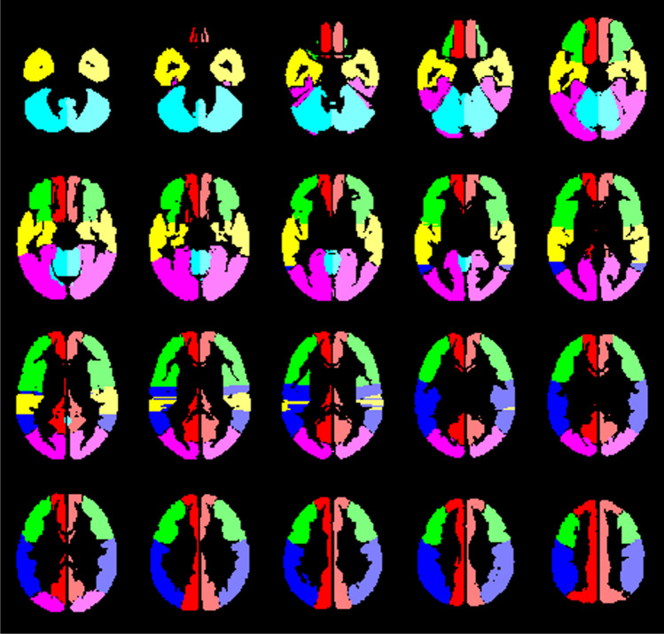
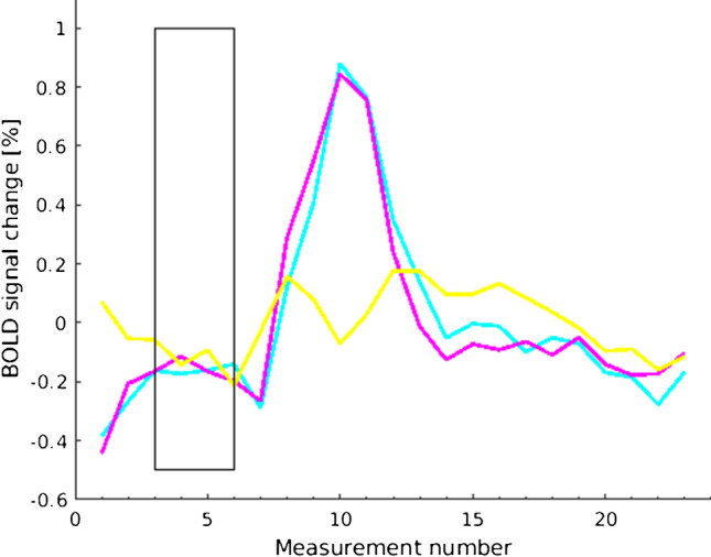
Methods: Patients with MMA underwent rs-fMRI and bh-fMRI in the same MRI session. Maps of the CVR gained by both modalities were compared retrospectively by calculating the correlation between the mean CVR of 12 volumes of interest. Additionally, the rs-maps of a subgroup of patients were compared to CPR-maps gained by [15O]water PET.

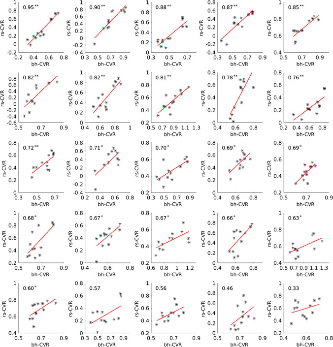
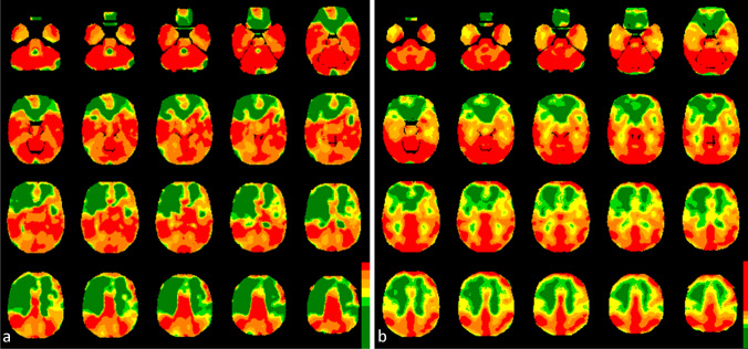
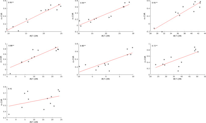
Results: The comparison of the rs-maps and the bh-maps of 24 patients revealed a good correlation (Pearson's r = 0.71 ± 0.13; preoperative patients: Pearson's r = 0.71 ± 0.17; postoperative patients: Pearson's r = 0.71 ± 0.11). The comparison of 7 rs-fMRI data sets to the corresponding [15O]water PET data sets also revealed a high level of agreement (Pearson's r = 0.80 ± 0.19).

Conclusion: The present analysis indicates that rs-fMRI might be a promising non-invasive method with almost no patient cooperation needed to evaluate the CVR. Further prospective studies are required.

Keywords: Breath-hold fMRI; Cerebrovascular reactivity; Moyamoya Angiopathy; Resting-state fMRI; [15O]water PET.


Conflict of interest statement

The authors declare that they have no conflict of interest.

Figures

Fig. 1
Fig. 1
The 12 evaluated volumes of interest based on the vascular territory of the anterior cerebral artery (red), the frontal (green), temporal (yellow), and parietal (blue) territories of the middle cerebral artery, the territory of the posterior cerebral artery (pink), and mainly the cerebellum/brainstem (turquoise), which were evaluated separately for both hemispheres
Fig. 2
Fig. 2
Exemplary BOLD signal time courses of one patient’s breath-hold measurement averaged over the 5 cycles. The rectangle marks the breath-hold period. Shown is a reference region (right cerebellum, turquoise), a region with physiological CVR (territory of the right posterior cerebral artery, pink), and a region with reduced CVR (temporal territory of the right middle cerebral artery, yellow)
Fig. 3
Fig. 3
Scatter plots showing the correlation between rs-fMRI CVR (correlation coefficients) and bh-fMRI CVR (mean relative signal change) of 12 VOIs of each patient’s measurement sorted by Pearson’s correlation coefficient in descending order. Two asterisks (**) mark significant correlation at p < 0.01. One asterisk (*) marks significant correlation at p < 0.05
Fig. 4
Fig. 4
CVR-maps of one patient (r = 0.90) gained by rs-fMRI (a) and bh-fMRI (b). The CVR values (rs-fMRI: correlation coefficients, bh-fMRI: mean relative signal change) are presented as overlays on normalized anatomical MRI images. Color-coding was applied with high CVR resembled by warm and low CVR by cold colors. The color-coding of both modalities was applied in standardized gradations using thresholds based on the individual 20th percentile of the cerebellar histograms (see “Methods” section). Both the CVR-maps gained by rs-fMRI and by bh-fMRI indicate reduced CVR in the territories of the anterior and the middle cerebral arteries whereat the right hemisphere is more affected than the left
Fig. 5
Fig. 5
Scatter plots showing the correlation between rs-fMRI CVR (correlation coefficients) and [15O]water PET CPR (mean cerebral blood flow changes (%) after ACZ stimulation) of 12 VOIs of each patient’s measurement sorted by Pearson’s correlation coefficient in descending order. Two asterisks (**) mark significant correlation at p < 0.01. One asterisk (*) marks significant correlation at p < 0.05
Fig. 6
Fig. 6
CVR-maps of one patient (r = 0.95) gained by rs-fMRI (a) and the corresponding CPR-maps gained by [15O]water PET with ACZ challenge (b). Color-coding was applied with high CVR/CPR resembled by warm and low CVR/CPR by cold colors. The rs-fMRI maps represent voxel-wise correlation coefficients to the cerebellum. All voxels exceeding the 20th percentile of the cerebellar histogram (correlation coefficient = 0.77) are colored red. The threshold values of the remaining colors result from a gradual subtraction of 0.2. Voxel-wise cerebral blood flow changes (%) after ACZ stimulation as provided by [15O]water PET are color-coded (modified “UCLA 2” color-scale, see “Methods” section). The maps of both modalities indicate perfusion deficits in the left hemisphere

References

    1. Suzuki J, Takaku A. Cerebrovascular “Moyamoya” disease: disease showing abnormal net-like vessels in base of brain. JAMA Neurol. 1969;20:288–299. - PubMed
    1. Kim JS. Moyamoya disease: epidemiology, clinical features, and diagnosis. J Stroke. 2016;18:2–11. - PMC - PubMed
    1. Scott RM, Smith ER. Moyamoya disease and moyamoya syndrome. N Engl J Med. 2009;360:1226–1237. - PubMed
    1. Tarasow E, Kulakowska A, Lukasiewicz A, et al. Moyamoya disease: diagnostic imaging. Pol J Radiol. 2011;76:73–79. - PMC - PubMed
    1. Khan N, Schuknecht B, Boltshauser E, et al. Moyamoya disease and Moyamoya syndrome: experience in Europe; choice of revascularisation procedures. Acta Neurochir (Wien) 2003;145:1061–1071. - PubMed