. 2021 Sep 27;16(9):e0248798.
doi: 10.1371/journal.pone.0248798. eCollection 2021.

Co-culture of type I and type II pneumocytes as a model of alveolar epithelium

Affiliations

Co-culture of type I and type II pneumocytes as a model of alveolar epithelium

Oliver Brookes et al. PLoS One. .

Abstract

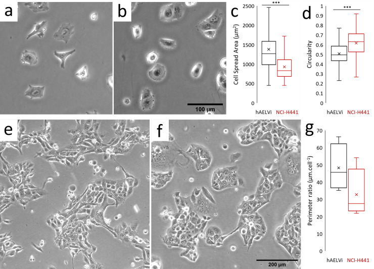
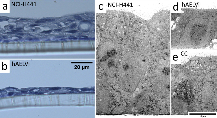
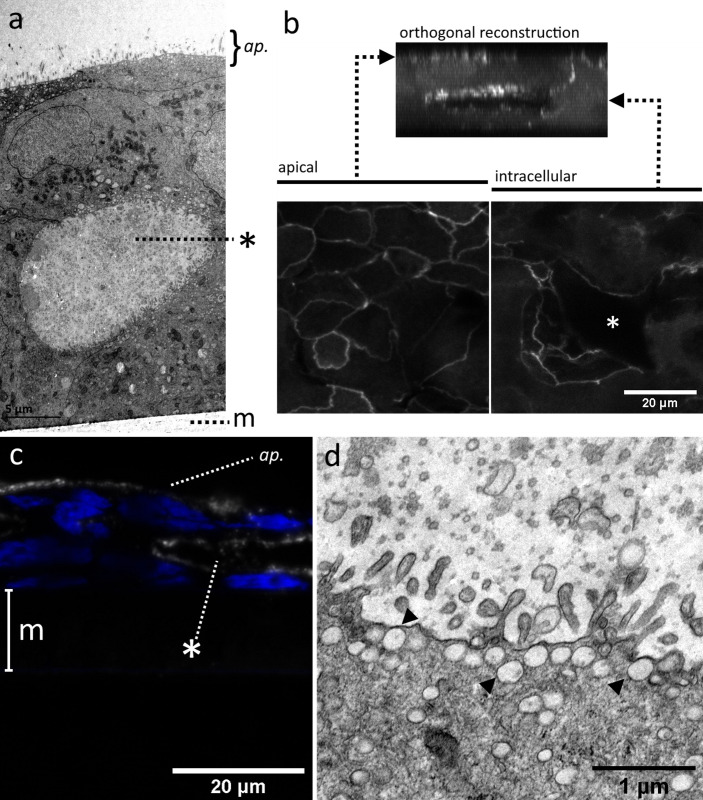
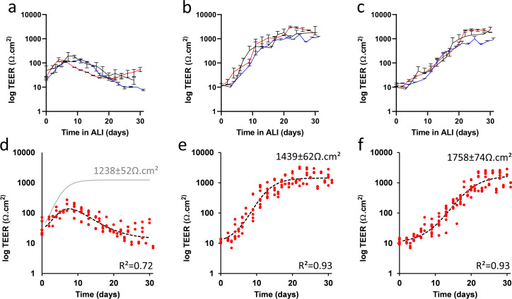
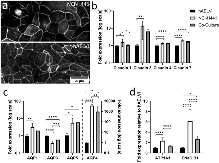
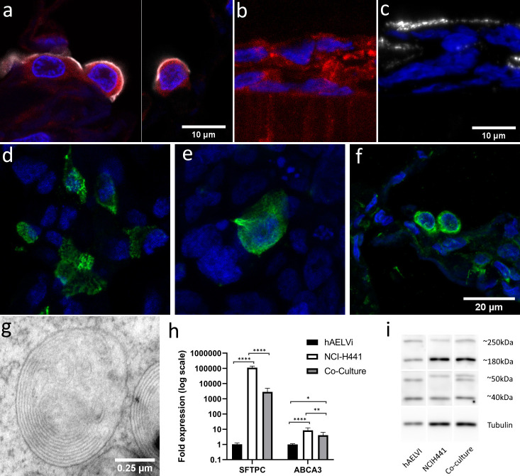
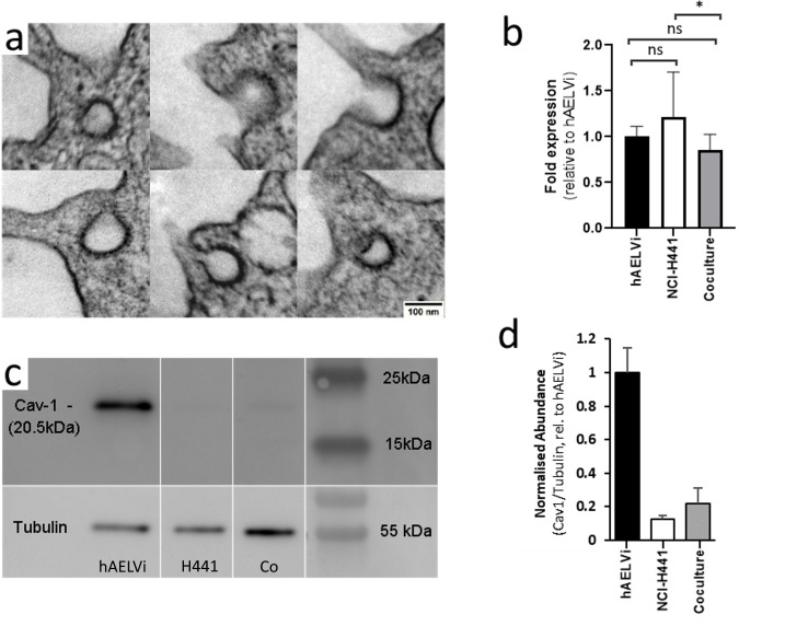
The epithelial tissues of the distal lung are continuously exposed to inhaled air, and are of research interest in studying respiratory exposure to both hazardous and therapeutic materials. Pharmaco-toxicological research depends on the development of sophisticated models of the alveolar epithelium, which better represent the different cell types present in the native lung and interactions between them. We developed an air-liquid interface (ALI) model of the alveolar epithelium which incorporates cell lines which bear features of type I (hAELVi) and type II (NCI-H441) epithelial cells. We compared morphology of single cells and the structure of cell layers of the two lines using light and electron microscopy. Working both in monotypic cultures and cocultures, we measured barrier function by trans-epithelial electrical resistance (TEER), and demonstrated that barrier properties can be maintained for 30 days. We created a mathematical model of TEER development over time based on these data in order to make inferences about the interactions occurring in these culture systems. We assessed expression of a panel of relevant genes that play important roles in barrier function and differentiation. The coculture model was observed to form a stable barrier akin to that seen in hAELVi, while expressing surfactant protein C, and having a profile of expression of claudins and aquaporins appropriate for the distal lung. We described cavities which arise within stratified cell layers in NCI-H441 and cocultured cells, and present evidence that these cavities represent an aberrant apical surface. In summary, our results support the coculture of these two cell lines to produce a model which better represents the breadth of functions seen in native alveolar epithelium.


Conflict of interest statement

The authors have declared that no competing interests exist.

Figures

Fig 1
Fig 1. Comparison of the morphology and growth habit of hAELVi and NCI-H441.
Phase contrast images of submerged cultures of hAELVi (a, e) and NCI-H441 (b, f) cells, at time points 6 hours (a, b) and 4 days after seeding on plastic culture flasks (e, f). Single cells were analysed within the captured images to determine cell spread area (c) and circularity (d) using ImageJ. Colony sizes were determined by measuring the length of the perimeters of colonies and normalising this value to the number of cells in the colony (g). *** = P<0.001, n = 15 or more cells in 5 or more randomly selected fields.
Fig 2
Fig 2. Supra-cellular structure of ALI cultures.
Light microscopy images of ultrathin sections stained with Papanicolaou stain (a, b) and TEM images (c, d, e) of ALI cultures of NCI-H441 (a, c) or hAELVi (b, d) or the coculture (e) after 14 days of culture. NCI-H441 cultures exhibit a loose stratified cell layer, while hAELVi typically form a flatter monolayer. Cocultures do exhibit stratification, but are generally more consistent than NCI-H441 alone.
Fig 3
Fig 3. TEM images of voids within stratified epithelial layers.
TEM images of NCI-H441:hAELVi cocultures grown at the air-liquid interface for 2 weeks reveal the existence of cavities beneath the surface (a). CLSM images of ZO-1 immunolabelled samples (b) indicate that these structures are lined with tight junctions on their luminal surface, in addition to those sealing the apical surface. Similar structures seen in formalin-fixed, paraffin embedded (FFPE) sections of NCI-H441 grown in ALI conditions stain positively using an antibody against HTII-280, an antigen found exclusively on the apical membrane of type II epithelial cells (c). Voids in NCI-H441 single cultures and cocultures are lined with microvillous processes and secretory vesicles, also features of the apical surface of type II alveolar epithelium (d). Asterisks mark internal voids. Polymer support membrane labelled m where visible. ap. = apical surface.
Fig 4
Fig 4. Electrophysiological barrier formation of NCI-H441, hAELVi, and co-culture in ALI conditions.
TEER measurements of NCI-H441 (a), hAELVi (b) and an equal co-culture of the two (c) over a period of thirty days following the establishment of ALI conditions. (3 independent experiments, n = 3 transwells). These data were fitted by regression to a logistic growth and decay model (d,e,f, red points indicate data values summarised in a,b and c, respectively). Only the model for NCI-H441 (d) includes a non-zero decay term; the dotted line shows the best fit including this decay term, while the grey line shows the logistic growth component in isolation for comparison to hAELVi (e) and co-culture (f). In preliminary experiments, we observed a direct mathematical relationship between TEER and Lucifer Yellow (LY) diffusion (S2 Fig in S1 File), suggesting that TEER and actual diffusion assays are mutually redundant, at least for LY, and that no new information is gained by performing both measurements.
Fig 5
Fig 5. Differences in expression of key functional genes between hAELVi, NCI-H441 and co-culture.
Cultures of hAELVi, NCI-H441 cells and a co-culture consisting of equal parts of both cell lines were maintained in ALI conditions for two weeks after confluence. Immunolabelling of ZO1 in the two parent lines revealed localisation to the cell periphery, consistent with incorporation into tight junctions (a). Gene expression analysis by RTqPCR of tight junction proteins (claudin 1, 2, 3 and 4; b), water channels (aquaporins AQP1, AQP3, AQP5 and AQP4 (c)) and ion pumps (ATP1A1 and ENaC B1; d) in hAELVi, NCI-H441 and a co-culture (fold expression relative to expression in hAELVi after normalisation to reference genes RPL13, RPL19 and HPRT. n = 3 replicates within 3 independent experiments.
Fig 6
Fig 6. NCI-H441 cells and co-cultures produce surfactant.
Immunofluorescent labelling of the lamellar body component ABCA3 (red) in normal human tissue samples (a) and in NCI-H441 ALI cultures (b). HTII-280, another type-II specific antigen (white) is present in the apical surface of type-II cells in normal human tissue (a) and in NCI-H441 (c) and cocultures after 14d (d). Immuno-labelling of pro-SPC reveals production of surfactant protein in cocultures (e) and normal human lung tissue (f). Electron microscopy reveals the presence of lamellar bodies beneath the surface of co-cultures of hAELVi and NCI-H441 cells grown in ALI conditions (g). RTqPCR reveals expression of both surfactant protein C and ABCA3 (h, n = 3 replicates within 3 independent experiments), and western blotting with an anti-ABCA3 polyclonal antibody reveals post-translational processing of ABCA3 (i).
Fig 7
Fig 7. Caveolae are observed in cocultures and Caveolin-1 is highly expressed in hAELVI.
Single cultures of NCI-H441 and hAELVi, and co-cultures consisting of equal proportions of both were seeded on permeable supports, cultured for two weeks at ALI and harvested. A portion of each membrane was fixed for TEM, and the remaining cells were lysed for extraction of RNA and protein. Structures bearing the ultrastructural features of caveolae were observed in some cells in co-culture (a). No caveoli were observed in NCI-H441 and hAELVi singly. Differences in CAV1 gene expression were not observed (b), but expression of Caveolin-1 protein is substantially higher in hAELVi than in NCIH-441 (c and d). n = 3 independent experiments, each containing minimum 3 technical replicates.

References

    1. Hynes J, Marshall L, Adcock I, Novotny T, Nic M, Dibusz K, et al.. Advanced Non-animal Models in Biomedical Research [Internet]. 2020. Available from: https://ec.europa.eu/jrc/en/publication/advanced-non-animal-models-biome...
    1. Sakagami M. In vitro, ex vivo and in vivo methods of lung absorption for inhaled drugs. Adv Drug Deliv Rev [Internet]. 2020; Available from: 10.1016/j.addr.2020.07.025 - DOI - PubMed
    1. Hermanns MI, Unger RE, Kehe K, Peters K, Kirkpatrick CJ. Lung epithelial cell lines in coculture with human pulmonary microvascular endothelial cells: Development of an alveolo-capillary barrier in vitro. Lab Investig. 2004;84(6):736–52. doi: 10.1038/labinvest.3700081 - DOI - PubMed
    1. Costa A, de Souza Carvalho-Wodarz C, Seabra V, Sarmento B, Lehr CM. Triple co-culture of human alveolar epithelium, endothelium and macrophages for studying the interaction of nanocarriers with the air-blood barrier. Acta Biomater [Internet]. 2019;91:235–47. Available from: 10.1016/j.actbio.2019.04.037 - DOI - PubMed
    1. Kletting S. Co-culture of human alveolar epithelial (hAELVi) and macrophage (THP-1) cell lines. ALTEX [Internet]. 2018;211–22. Available from: https://www.altex.org/index.php/altex/article/view/89 doi: 10.14573/altex.1607191 - DOI - PubMed

Publication types