. 2022 Jan;94(1):99-109.
doi: 10.1002/jmv.27358. Epub 2021 Oct 5.

A systematic review on SARS-CoV-2-associated fungal coinfections

Affiliations

A systematic review on SARS-CoV-2-associated fungal coinfections

Shringika Soni et al. J Med Virol. 2022 Jan.

Abstract

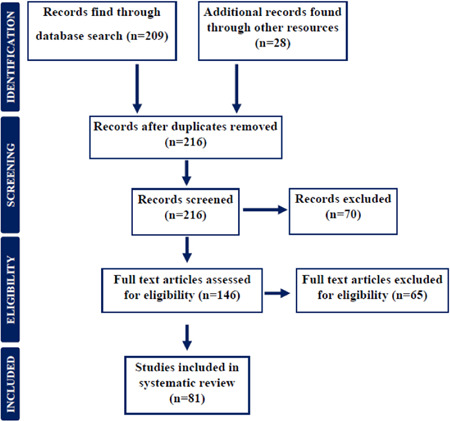
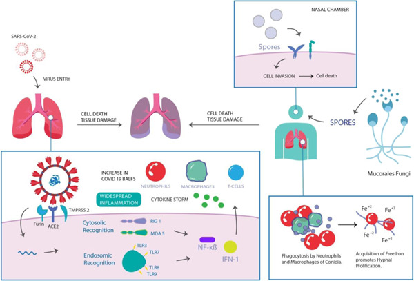
A severe pandemic of Coronavirus Disease (COVID-19) has been sweeping the globe since 2019, and this time, it did not stop, with frequent mutations transforming into virulent strains, for instance, B.1.1.7, B.1.351, and B.1.427. In recent months, a fungal infection, mucormycosis has emerged with more fatal responses and significantly increased mortality rate. To measure the severity and potential alternative approaches against black fungus coinfection in COVID-19 patients, PubMed, Google Scholar, World Health Organization (WHO) newsletters, and other online resources, based on the cases reported and retrospective observational analysis were searched from the years 2015-2021. The studies reporting mucormycosis with Severe Acute Respiratory Syndrome Coronavirus 2 (SARS-CoV-2) coinfection and/or demonstrating potential risk factors, such as a history of diabetes mellitus or suppressed immune system were included, and reports published in non-English language were excluded. More than 20 case reports and observational studies on black fungus coinfection in COVID-19 patients were eligible for inclusion. The results indicated that diabetes mellitus, hyperglycemic, and immunocompromised COVID-19 patients with mucormycosis were at a higher risk. We found that it was prudent to assess the potential risk factors and severity of invasive mycosis via standardized diagnostic and clinical settings. Large-scale studies need to be conducted to identify early biomarkers and optimization of diagnostic methods has to be established per population and geographical variation. This will not only help clinicians around the world to detect the coinfection in time but also will prepare them for future outbreaks of other potential pandemics.

Keywords: COVID-19; black fungus; diagnosis; fungal infection; mucormycosis.


Conflict of interest statement

The authors declare that there are no conflict of interests.

Figures

Figure 1
Figure 1
Preferred reporting flow chart for including and studying studies and case reports for systematic review
Figure 2
Figure 2
Representation of the potential association between SARS‐CoV‐2 and mucormycosis
Figure 3
Figure 3
Identification of mucormycosis in COVID‐19 patients: (A), (B) Positron Emission Tomography (PET)‐Computed tomography (CT) imaging demonstrating nodule in right upper lobe posterior segment along with cavitating necrosis. (C), (D) Hypermetabolic mediastinal nodes with soft tissue nodules in right arms and thigh. (E) Nonseptate fungal hyphae, suggestive of mucormycosis via microscopy/histopathological imaging representation. Reproduced from (69), Copyright 2021, with permission from IP Indian Journal of Immunology and Respiratory Medicine. COVID‐19, coronavirus disease 2019
Figure 4
Figure 4
Post‐COVID mucormycosis radiographic imaging presenting: (A−C) Computed tomography (CT) scans of intracranial involvement in paranasal sinuses, ethmoid group. (D−I) Axial, magnetic resonance imaging of mucormycosis induced intra‐orbital extension in post‐COVID patients. Reproduced from (33), Copyright 2021, with permission Cambridge Press.COVID, coronavirus disease
Figure 5
Figure 5
(A) Computed tomography (CT) representation of peripheral ground‐glass opacities in both lungs of mucormycosis in SARS‐CoV‐2 coinfection. (B) Magnetic resonance imaging (MRI) imaging of coronal section (T1) presenting irregular hypersensitivity of retrobulbar space in COVID‐19 patient, which suggested the presence of fungal infection. Reproduced from (74), Copyright 2020, with permission Cureus

Similar articles

Cited by

References

    1. Mallapaty S. What's the risk of dying from a fast‐spreading COVID‐19 variant? Nature. 2021;590(7845):191‐192. - PubMed
    1. Thiagarajan K. Why is India having a covid‐19 surge? BMJ. 2021;373:n1124. - PubMed
    1. Pemán J, Ruiz‐Gaitán A, García‐Vidal C, et al. Fungal co‐infection in COVID‐19 patients: should we be concerned? Rev Iberoam Micol. 2020;37(2):41‐46. - PMC - PubMed
    1. Allegra A, Di Gioacchino M, Tonacci A, Musolino C, Gangemi S. Immunopathology of SARS‐CoV‐2 infection: immune cells and mediators, prognostic factors, and immune‐therapeutic implications. Int J Mol Sci. 2020;21(13):4782. - PMC - PubMed
    1. Chauhan N, Soni S, Jain U. Optimizing testing regimes for the detection of COVID‐19 in children and older adults. Expert Rev Mol Diagn. 2021:1‐18. 10.1080/14737159.2021.1962708 - DOI - PMC - PubMed

Publication types

MeSH terms