. 2021 Sep 18;10(9):931.
doi: 10.3390/biology10090931.

Human Periodontal Ligament Stem Cells Response to Titanium Implant Surface: Extracellular Matrix Deposition

Affiliations

Human Periodontal Ligament Stem Cells Response to Titanium Implant Surface: Extracellular Matrix Deposition

Guya Diletta Marconi et al. Biology (Basel). .

Erratum in

Abstract

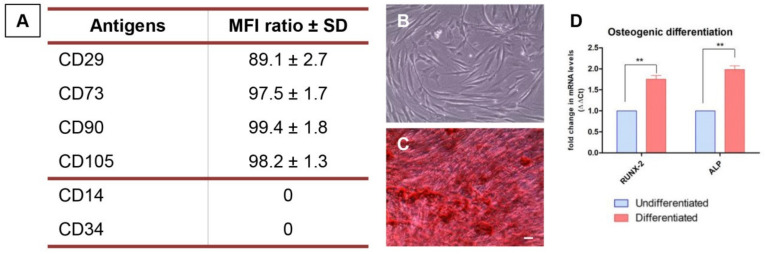
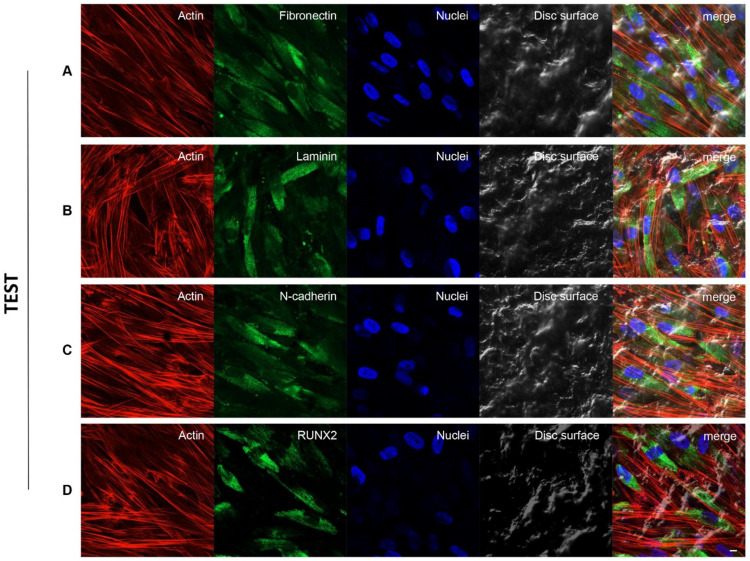
The major challenge for dentistry is to provide the patient an oral rehabilitation to maintain healthy bone conditions in order to reduce the time for loading protocols. Advancement in implant surface design is necessary to favour and promote the osseointegration process. The surface features of titanium dental implant can promote a relevant influence on the morphology and differentiation ability of mesenchymal stem cells, induction of the osteoblastic genes expression and the release of extracellular matrix (ECM) components. The present study aimed at evaluating the in vitro effects of two different dental implants with titanium surfaces, TEST and CTRL, to culture the human periodontal ligament stem cells (hPDLSCs). Expression of ECM components such as Vimentin, Fibronectin, N-cadherin, Laminin, Focal Adhesion Kinase (FAK) and Integrin beta-1 (ITGB1), and the osteogenic related markers, as runt related transcription factor 2 (RUNX2) and alkaline phosphatase (ALP), were investigated. Human PDLSCs cultured on the TEST implant surface demonstrated a better cell adhesion capability as observed by Scanning Electron Microscopy (SEM) and immunofluorescence analysis. Moreover, immunofluorescence and Western blot experiments showed an over expression of Fibronectin, Laminin, N-cadherin and RUNX2 in hPDLSCs seeded on TEST implant surface. The gene expression study by RT-PCR validated the results obtained in protein assays and exhibited the expression of RUNX2, ALP, Vimentin (VIM), Fibronectin (FN1), N-cadherin (CDH2), Laminin (LAMB1), FAK and ITGB1 in hPDLSCs seeded on TEST surface compared to the CTRL dental implant surface. Understanding the mechanisms of ECM components release and its regulation are essential for developing novel strategies in tissue engineering and regenerative medicine. Our results demonstrated that the impact of treated surfaces of titanium dental implants might increase and accelerate the ECM apposition and provide the starting point to initiate the osseointegration process.

Keywords: adhesion; extracellular matrix; gene expression; osseointegration; osteogenesis.


Conflict of interest statement

The authors declare no conflict of interest.

Figures

Figure 1
Figure 1
hPDLSCs characterization. (A) Flow cytometry results of the expression of surface markers. (B) Representative light microscopy image of plastic-adherent hPDLSCs cultured in standard conditions. (C) Cells were positive for Alizarin red S staining after 3 weeks of differentiation. (D) RT-PCR showed the upregulation of osteogenic related in differentiated hPDLSCs. Scale bar: 10 μm. ** p < 0.01.
Figure 2
Figure 2
(A) SEM observations of CTRL surface without cells captured at 50× magnification. (B) SEM observations of CTRL surface without cells captured at 1000× magnification. (C) SEM observations of hPDLSCs cultured on CTRL surface captured at 1000× magnification. (D1D4) CLSM observation of hPDLSCs cultured on CTRL surface. (D1) Cytoskeleton actin stained with Alexa Fluor 594 phalloidin and observed under red fluorescence channel; (D2) Nuclei stained with TOPRO and observed under blue fluorescence channel; (D3) CTRL surface observed under light transmission channel; (D4) merged image of above mentioned channels. Scale bar: 20 μm. White arrows indicate adherent cells on CTRL surface.
Figure 3
Figure 3
(A) SEM observations of TEST surface without cells captured at 50× magnification. (B) SEM observations of TEST surface without cells captured at 1000× magnification. (C) SEM observations of hPDLSCs cultured on TEST surface captured at 1000× magnification. (D1–D4) CLSM observation of hPDLSCs cultured on TEST surface. (D1) Cytoskeleton actin stained with Alexa Fluor 594 phalloidin and observed under red fluorescence channel; (D2) Nuclei stained with TOPRO and observed under blue fluorescence channel; (D3) TEST surface observed under light transmission channel; (D4) merged image of above mentioned channels. Scale bar: 20 μm. White arrows indicate adherent cells on TEST surface.
Figure 4
Figure 4
Human PDLSCs cultured on CTRL titanium implant surface were observed after 8 weeks of culture. Cytoskeleton actin was stained in red fluorescence; specific markers (A) Fibronectin, (B) Laminin, (C) N-cadherin and (D) RUNX2, were stained in green fluorescence; nuclei were stained in blue fluorescence. Scale bar: 10 µm.
Figure 5
Figure 5
Human PDLSCs cultured on TEST titanium implant surface were observed after 8 weeks of incubation. Cytoskeleton actin was stained in red fluorescence; specific markers (A) Fibronectin, (B) Laminin, (C) N-cadherin and (D) RUNX2, were stained in green fluorescence; nuclei were stained in blue fluorescence. Scale bar: 10 µm.
Figure 6
Figure 6
(A) Graph of RT-PCR showed the mRNA levels of RUNX2, ALP, VIM, FN1, CDH2, LAMB1, FAK and ITGB1 in cells cultured on CTRL and TEST surface. * p < 0.05; ** p < 0.01. (B) Protein level expression of Fibronectin, Laminin, N-cadherin and RUNX2 in cells cultured on CTRL and TEST surface. β-actin was used as a housekeeping protein. Graph bars represent the densitometric measurements of proteins bands expressed as integrated optical intensity with the mean of three separate experiments. The error bars in these graphs showed the standard deviation (±SD). Densitometric values analysed by ANOVA showed significant differences. ** p < 0.01; *** p < 0.001. Please refer to Full Western blot in Supplementary.

Similar articles

Cited by

References

    1. Le Guehennec L., Soueidan A., Layrolle P., Amouriq Y. Surface treatments of titanium dental implants for rapid osseointegration. Dent. Mater. 2007;23:844–854. doi: 10.1016/j.dental.2006.06.025. - DOI - PubMed
    1. Attanasio F., Antonelli A., Brancaccio Y., Averta F., Figliuzzi M.M., Fortunato L., Giudice A. Primary Stability of Three Different Osteotomy Techniques in Medullary Bone: An in Vitro Study. Dent. J. 2020;8:21. doi: 10.3390/dj8010021. - DOI - PMC - PubMed
    1. Liddell R.S., Ajami E., Li Y., Bajenova E., Yang Y., Davies J.E. The influence of implant design on the kinetics of osseointegration and bone anchorage homeostasis. Acta Biomater. 2021;121:514–526. doi: 10.1016/j.actbio.2020.11.043. - DOI - PubMed
    1. Sakka S., Coulthard P. Bone quality: A reality for the process of osseointegration. Implant. Dent. 2009;18:480–485. doi: 10.1097/ID.0b013e3181bb840d. - DOI - PubMed
    1. Bosshardt D.D., Chappuis V., Buser D. Osseointegration of titanium, titanium alloy and zirconia dental implants: Current knowledge and open questions. Periodontol. 2000. 2017;73:22–40. doi: 10.1111/prd.12179. - DOI - PubMed