. 2021 Sep 19;10(9):936.
doi: 10.3390/biology10090936.

Identification and Characterization of a Rhodopsin Kinase Gene in the Suckers of Octopus vulgaris: Looking around Using Arms?

Affiliations

Identification and Characterization of a Rhodopsin Kinase Gene in the Suckers of Octopus vulgaris: Looking around Using Arms?

Al-Sayed Al-Soudy et al. Biology (Basel). .

Abstract

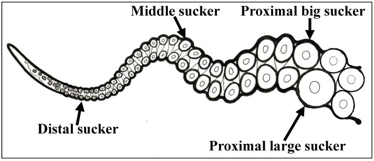
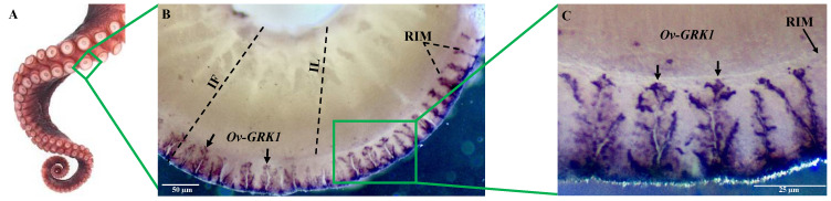
In their foraging behavior octopuses rely on arm search movements outside the visual field of the eyes. In these movements the environment is explored primarily by the suckers that line the entire length of the octopus arm. In this study, for the first time, we report the complete characterization of a light-sensing molecule, Ov-GRK1, in the suckers, skin and retina of Octopus vulgaris. We sequenced the O. vulgaris GRK1 gene, defining a phylogenetic tree and performing a 3D structure model prediction. Furthermore, we found differences in relative mRNA expression in different sucker types at several arm levels, and localized it through in situ hybridization. Our findings suggest that the suckers in octopus arms are much more multimodal than was previously shown, adding the potential for light sensing to the already known mechanical and chemical sensing abilities.

Keywords: GRK1; arm suckers; cephalopods; extra-ocular perception; octopus; retina; skin.


Conflict of interest statement

The authors declare no conflict of interest.

Figures

Figure 1
Figure 1
Image description of four sucker types: proximal big, proximal large, middle, distal.
Figure 2
Figure 2
Gene expression analysis for mRNA of Rhodopsin kinase receptor gene (Ov-GRK1) of different tissues. Relative mRNA expression levels were measured using real-time analysis and calculated by the 2(−ΔΔC(T)) method. Each sample (n = 3) was tested and run in duplicate. The heart tissue was used as a negative control. The ubiquitin gene was amplified as an internal control. No-template controls were included. Relative mRNA fold change in gene expression was compared to the proximal big sucker (set y = 1). * asterisk indicates that the difference vs. sucker proximal big is statistically significant (Wilcoxon-test, p < 0.05). The Kruskal–Wallis test showed that there was a statistically significance between groups (p < 0.05). Error bars represent the SEM.
Figure 3
Figure 3
Whole-mount in situ hybridization of the Ov-GRK1 gene in the rim of an O. vulgaris middle sucker. (A) Octopus arm with array of suckers; (B) the receptor expression is present in the epithelium of the rim (arrows); (C) higher magnification of the portion of the epithelium rim inside the green rectangle showing Ov-GRK1 expression. Sucker rim epithelium (RIM), sucker infundibulum (IF), infundibulum lumen (IL). Arrows indicate positive signals.
Figure 4
Figure 4
GRK1 amino acid sequences alignment. Alignment of Ov-GRK1 amino acid sequences to an O. bimaculoides rhodopsin kinase amino acid sequence from GenBank (accession number XP014774259) and to those of E. scolopes rhodopsin kinase extracted from the eyes (ACB05676) and light organ (ACB05677). White line boxes highlight potential amino acid differences among samples. Complete open reading frames are shown.
Figure 5
Figure 5
Bayesian tree of GRK protein sequences. Bayesian phylogenetic tree performed with LG + G model, constructed with protein sequences from Ov-GRK1 and Rhodopsin kinase receptors of 18 species including other cephalopods, invertebrates and vertebrates. All protein sequences were obtained from GenBank and the accession numbers are presented in Table S1. Bayesian posterior probabilities are represented over nodes; turquoise box highlights the cephalopod clade and the Ov-GRK1 protein sequence is in bold.
Figure 6
Figure 6
Homology modeling of the Ov-GRK1. (A) Alignment with a potential Model-Template (6c2y). (B) 3D structural view for the prediction of ligand binding sites in Ov-GRK1 generated by 3DLigandSite based on the PDB.

References

    1. Hanlon R., Messenger J. Adaptive coloration in young cuttlefish (Sepia officinalis L.): The morphology and development of body patterns and their relation to behavior. Philos. Trans. R. Soc. B Biol. Sci. 1988;320:437–487.
    1. Chiao C.-C., Chubb C., Buresch K.C., Barbosa A., Allen J.J., Mäthger L.M., Hanlon R.T. Mottle camouflage patterns in cuttlefish: Quantitative characterization and visual background stimuli that evoke them. J. Exp. Biol. 2010;213:187–199. doi: 10.1242/jeb.030247. - DOI - PubMed
    1. Cloney R., Brocco S. Chromatophore organs, reflector cells, iridocytes and leucophores in cephalopods. Am. Zool. 1983;23:581–892. doi: 10.1093/icb/23.3.581. - DOI
    1. Allen J.J., Mäthger L.M., Barbosa A., Hanlon R.T. Cuttlefish use visual cues to control three-dimensional skin papillae for camouflage. J. Comp. Physiol. A Neuroethol. Sens. Neural Behav. Physiol. 2009;195:547–555. doi: 10.1007/s00359-009-0430-y. - DOI - PubMed
    1. Romagny S., Darmaillacq A.S., Guibé M., Bellanger C., Dickel L. Feel, smell and see in an egg: Emergence of perception and learning in an immature invertebrate, the cuttlefish embryo. J. Exp. Biol. 2012;215:4125–4130. doi: 10.1242/jeb.078295. - DOI - PubMed

LinkOut - more resources