Review
. 2021 Aug 25;10(9):2198.
doi: 10.3390/cells10092198.

Down the Iron Path: Mitochondrial Iron Homeostasis and Beyond

Affiliations
Review

Down the Iron Path: Mitochondrial Iron Homeostasis and Beyond

Jonathan V Dietz et al. Cells. .

Abstract

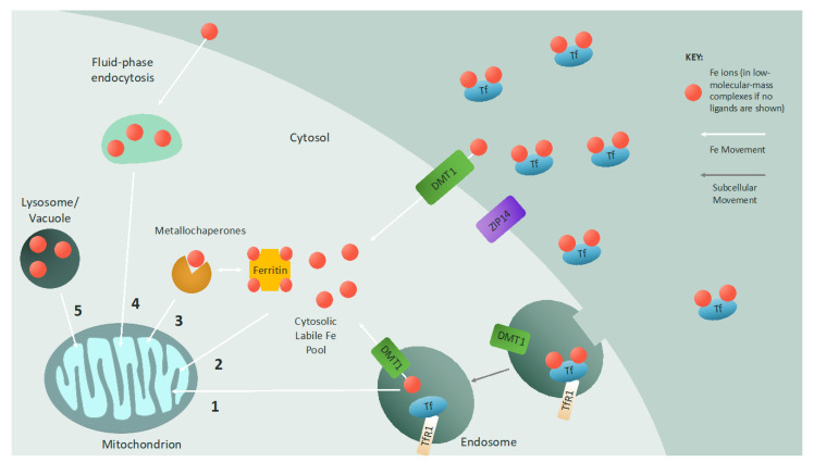
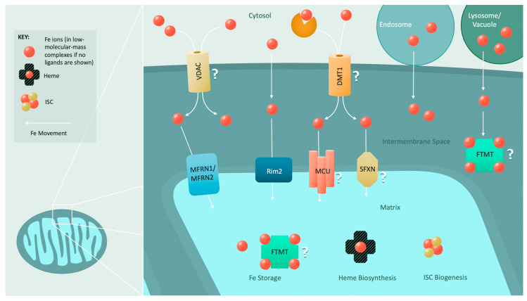
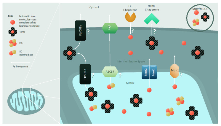
Cellular iron homeostasis and mitochondrial iron homeostasis are interdependent. Mitochondria must import iron to form iron-sulfur clusters and heme, and to incorporate these cofactors along with iron ions into mitochondrial proteins that support essential functions, including cellular respiration. In turn, mitochondria supply the cell with heme and enable the biogenesis of cytosolic and nuclear proteins containing iron-sulfur clusters. Impairment in cellular or mitochondrial iron homeostasis is deleterious and can result in numerous human diseases. Due to its reactivity, iron is stored and trafficked through the body, intracellularly, and within mitochondria via carefully orchestrated processes. Here, we focus on describing the processes of and components involved in mitochondrial iron trafficking and storage, as well as mitochondrial iron-sulfur cluster biogenesis and heme biosynthesis. Recent findings and the most pressing topics for future research are highlighted.

Keywords: heme biosynthesis; iron homeostasis; iron trafficking; mitochondrial iron–sulfur clusters.


Conflict of interest statement

The authors declare no conflict of interest. The funders had no role in the interpretation of data or in the writing of the manuscript.

Figures

Figure 1
Figure 1
Iron delivery to the mitochondrion: Iron is imported into cells by either endocytosis (transferrin-bound iron) or plasma membrane channels (non-transferrin-bound iron). The metal transporter ZIP14 has been shown to aid in the cellular uptake of both transferrin-bound and non-transferrin-bound iron. When iron-loaded transferrin binds to the transferrin receptor on the cell surface, the protein–receptor complex is endocytosed, and iron is released into the endosome and reduced. This iron can be exported via the endosomal divalent metal transporter (DMT1) to the cytosolic labile iron pool, which consists of iron ions in low-molecular-mass complexes or stored in ferritin. Alternatively, if the endosome contacts the mitochondrion, iron is transferred to the mitochondrion via a “kiss and run” mechanism (1). Non-transferrin-bound iron imported by DMT1 on the cell membrane directly enters the cytosolic labile iron pool as iron chelated by low-molecular-mass molecules or ferritin and can then be imported into mitochondria (2). Metallochaperones such as glutaredoxin can also deliver iron to mitochondria (3). Fluid-phase endocytosis is an additional mechanism to deliver non-transferrin-bound iron to mitochondria (4). In some cases, interactions between mitochondria and lysosomes or vacuoles mediate iron transfer between organelles (5), and there may be a synergistic effect between lysosome/vacuole homeostasis and mitochondrial iron homeostasis. (Tf = transferrin; TfR1 = transferrin receptor 1).
Figure 2
Figure 2
Mitochondrial iron import and utilization: The mechanism for iron import through the outer mitochondrial membrane is poorly understood. However, it is speculated that ion channels such as VDACs and a recently identified mitochondrial divalent metal transporter (DMT1) facilitate the import of iron. Interorganellar contacts with endosomes, lysosomes, and vacuoles can also convey iron to mitochondria. Transport across the inner mitochondrial membrane is primarily driven by the conserved mitoferrins (MFRN1 and MFRN2). In yeast, a low-affinity iron transporter (Rim2) assists the mitoferrins. Roles in inner mitochondrial membrane iron import have also been hypothesized for the mitochondrial calcium uniporter (MCU) and several members of the sideroflexin (SFXN) family. Once iron is imported into the mitochondrion, it is utilized for heme biosynthesis or ISC biogenesis, or it is stored in mitochondrial ferritin (FTMT) or within low-molecular-mass complexes.
Figure 3
Figure 3
Export of mitochondrial iron and iron-containing cofactors: Iron is exported from mitochondria as heme or metal ions in low-molecular-mass complexes. Additionally, a presently unidentified intermediate required for cytosolic ISC biogenesis is also exported from mitochondria. In yeast, three inner mitochondrial membrane iron exporters have been identified: Mmt1, Mmt2, and Mtm1. In the case of heme export, FLVCR1b is postulated to play a role, but it is unclear where FLVCR1b localizes within the mitochondrion or to what extent it facilitates heme export. The ISC intermediate necessary for cytosolic ISC biogenesis is trafficked across the inner mitochondrial membrane by Atm1 in yeast and ABCB7 in mammals. Methods of export across the outer mitochondrial membrane remain unknown. It is hypothesized that various protein channels allow for the flow of these iron species, and that iron and cofactor chaperones directly accept or extract their cargo from the mitochondrion. A non-protein-based mechanism for iron, heme, and ISC intermediate export is also possible via mitochondria-derived vesicles (MDVs) and compartments (MDCs).

References

    1. Gao J., Zhou Q., Wu D., Chen L. Mitochondrial iron metabolism and its role in diseases. Clin. Chim. Acta. 2020;513:6–12. doi: 10.1016/j.cca.2020.12.005. - DOI - PubMed
    1. Puig S., Askeland E., Thiele D.J. Coordinated remodeling of cellular metabolism during iron deficiency through targeted mRNA degradation. Cell. 2005;120:99–110. doi: 10.1016/j.cell.2004.11.032. - DOI - PubMed
    1. Siekevitz P. Powerhouse of the cell. Sci. Am. 1957;197:131–144. doi: 10.1038/scientificamerican0757-131. - DOI
    1. Van der Bliek A., Sedensky M.M., Morgan P.G. Cell biology of the mitochondrion. Genetics. 2017;207:843–871. doi: 10.1534/genetics.117.300262. - DOI - PMC - PubMed
    1. Cheng Z., Ristow M. Mitochondria and metabolic homeostasis. Antioxid. Redox Signal. 2013;19:240–242. doi: 10.1089/ars.2013.5255. - DOI - PubMed

Publication types