Review
. 2021 Aug 30;10(9):2245.
doi: 10.3390/cells10092245.

Annexin A1 as a Regulator of Immune Response in Cancer

Affiliations
Review

Annexin A1 as a Regulator of Immune Response in Cancer

Thaise Gonçalves Araújo et al. Cells. .

Abstract

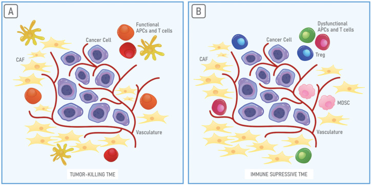
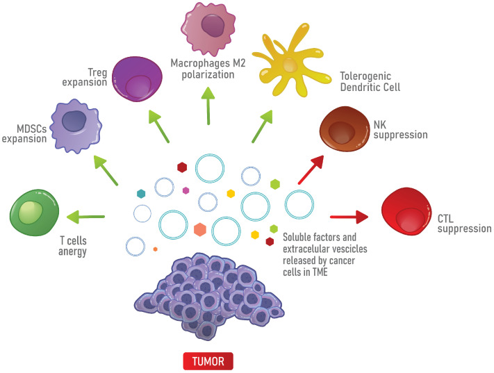
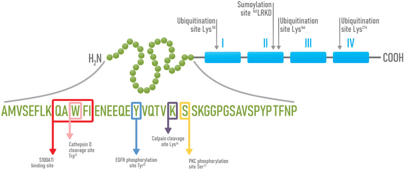
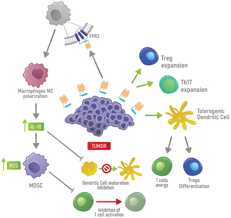
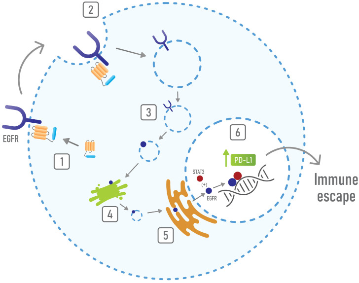
Annexin A1 is a 37 kDa phospholipid-binding protein that is expressed in many tissues and cell types, including leukocytes, lymphocytes and epithelial cells. Although Annexin A1 has been extensively studied for its anti-inflammatory activity, it has been shown that, in the cancer context, its activity switches from anti-inflammatory to pro-inflammatory. Remarkably, Annexin A1 shows pro-invasive and pro-tumoral properties in several cancers either by eliciting autocrine signaling in cancer cells or by inducing a favorable tumor microenvironment. Indeed, the signaling of the N-terminal peptide of AnxA1 has been described to promote the switching of macrophages to the pro-tumoral M2 phenotype. Moreover, AnxA1 has been described to prevent the induction of antigen-specific cytotoxic T cell response and to play an essential role in the induction of regulatory T lymphocytes. In this way, Annexin A1 inhibits the anti-tumor immunity and supports the formation of an immunosuppressed tumor microenvironment that promotes tumor growth and metastasis. For these reasons, in this review we aim to describe the role of Annexin A1 in the establishment of the tumor microenvironment, focusing on the immunosuppressive and immunomodulatory activities of Annexin A1 and on its interaction with the epidermal growth factor receptor.

Keywords: Annexin A1; cancer aggressiveness; immune-suppression; tumor microenvironment.


Conflict of interest statement

The authors declare no conflict of interest. The funders had no role in the design of the study; in the collection, analyses, or interpretation of data; in the writing of the manuscript, or in the decision to publish the results.

Figures

Figure 1
Figure 1
The tumor microenvironment (TME). Cancer cells are surrounded by a complex and heterogeneous environment composed of cancer-associated fibroblasts (CAFs), blood vessel cells and immune system cells, the TME. (A) Whether enriched in functional antigen-presenting cells (APCs) or T cells, the TME can promote the recognition of cancer cells by the immune system. (B) On the contrary, the TME can be enriched in dysfunctional APCs and T cells, myeloid-derived suppressor cells (MDSCs) and Tregs, creating a highly immune suppressive environment that favors tumor growth and progression.
Figure 2
Figure 2
Immune suppression in the TME. In the TME, soluble factors and extracellular vesicles (EVs) released by cancer cells promote the differentiation into immune suppressive cells, such as Tregs, MDSCs, M2 macrophages and tolerogenic DCs, which in turn, inhibit the anti-tumor immune response mediated by T cells, CTLs and NK cells.
Figure 3
Figure 3
Schematic representation of AnxA1 structure and its sites of post-translational modifications. AnxA1 is composed of an N-terminal peptide (green) and a C-terminal region, composed of four repetitive domains of about 70 aa each (blue). In the N-terminal domain, AnxA1 display sites for EGFR (Y, Tyr21) and PKC dependent phosphorylation (S, Ser27), sites for Cathepsin D (W, Trp12) and Calpain I dependent cleavages (K, Lys26) and a peptide motif (QAWFI), responsible for the binding to S100A11. In the C-terminal portion, three sites of ubiquitination (Lys58, Lys166 and Lys276) and a site of sumoylation (160 LRKD) are present.
Figure 4
Figure 4
Mechanism of immune escape induced by AnxA1 in the TME. AnxA1 expressed by cancer cells promotes the expansion of Tregs, tDCs and TH17. Moreover, AnxA1 can activate FPR2 on the cell surface of macrophages, supporting the polarization toward an M2 phenotype. In turn, M2 macrophages induce MDSC activity and inhibit the maturation of DCs by producing IL-10.
Figure 5
Figure 5
Mechanisms of immune escape mediated by the AnxA1/EGFR interaction. AnxA1 interacts with EGFR (1) and promotes its internalization (2 and 3) and its retrograde transport to nuclei (4 and 5). Once in nuclei, EGFR interacts with STAT3 and induces the expression of PD-L1 (6), thus resulting in immune suppression.

References

    1. Hanahan D., Weinberg R.A. Hallmarks of cancer: The next generation. Cell. 2011;144:646–674. doi: 10.1016/j.cell.2011.02.013. - DOI - PubMed
    1. Brassart-Pasco S., Brezillon S., Brassart B., Ramont L., Oudart J.B., Monboisse J.C. Tumor Microenvironment: Extracellular Matrix Alterations Influence Tumor Progression. Front. Oncol. 2020;10:397. doi: 10.3389/fonc.2020.00397. - DOI - PMC - PubMed
    1. Chaffer C.L., Weinberg R.A. A perspective on cancer cell metastasis. Science. 2011;331:1559–1564. doi: 10.1126/science.1203543. - DOI - PubMed
    1. Blankenstein T., Coulie P.G., Gilboa E., Jaffee E.M. The determinants of tumour immunogenicity. Nat. Rev. Cancer. 2012;12:307–313. doi: 10.1038/nrc3246. - DOI - PMC - PubMed
    1. Pradeu T., Carosella E.D. On the definition of a criterion of immunogenicity. Proc. Natl. Acad. Sci. USA. 2006;103:17858–17861. doi: 10.1073/pnas.0608683103. - DOI - PMC - PubMed

Publication types