Review
. 2021 Aug 31;10(9):2254.
doi: 10.3390/cells10092254.

p27Kip1, an Intrinsically Unstructured Protein with Scaffold Properties

Affiliations
Review

p27Kip1, an Intrinsically Unstructured Protein with Scaffold Properties

Debora Bencivenga et al. Cells. .

Abstract

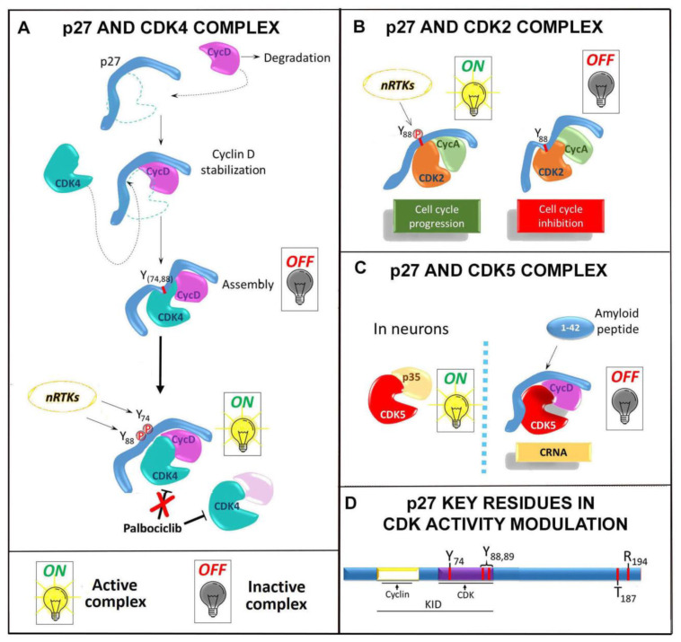
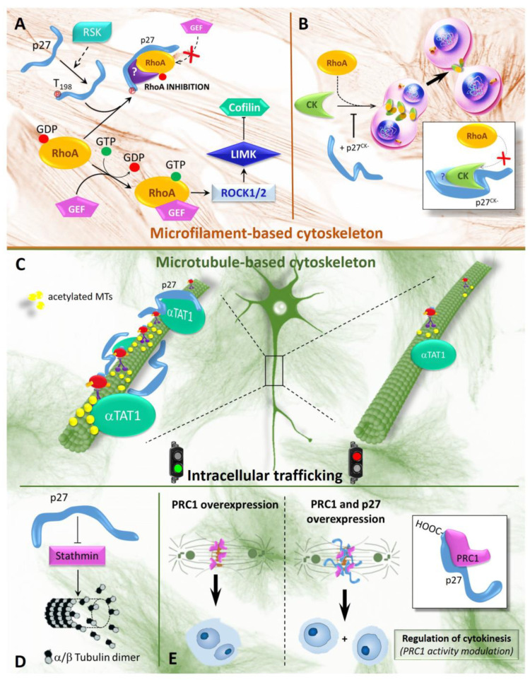
The Cyclin-dependent kinase (CDK) regulator p27Kip1 is a gatekeeper of G1/S transition. It also regulates G2/M progression and cytokinesis completion, via CDK-dependent or -independent mechanisms. Recently, other important p27Kip1 functions have been described, including the regulation of cell motility and migration, the control of cell differentiation program and the activation of apoptosis/autophagy. Several factors modulate p27Kip1 activities, including its level, cellular localization and post-translational modifications. As a matter of fact, the protein is phosphorylated, ubiquitinated, SUMOylated, O-linked N-acetylglicosylated and acetylated on different residues. p27Kip1 belongs to the family of the intrinsically unstructured proteins and thus it is endowed with a large flexibility and numerous interactors, only partially identified. In this review, we look at p27Kip1 properties and ascribe part of its heterogeneous functions to the ability to act as an anchor or scaffold capable to participate in the construction of different platforms for modulating cell response to extracellular signals and allowing adaptation to environmental changes.

Keywords: CDK; Rho GTPase; cyclin; cytoskeleton; intrinsically unstructured protein; p27Kip1; scaffold protein; αTAT1.


Conflict of interest statement

The authors declare no conflict of interest.

Figures

Figure 1
Figure 1
p27 and CDK interaction. The scheme summarizes some of p27 interaction with different Cyclin/CDK heterodimers. The sequence of the events is reported in detail in the text. (A) p27 binds sequentially Cyclin D and CDK4, facilitating the formation of the kinase complex. The modification of Tyrosine 74 (88) (P in red) by non-receptor tyrosine kinases (nRTKs) weakens the affinity of p27 for CDK4 which is then free to allocate the ATP in its specific ATP-binding site, allowing the full activation of the kinase complex. When bound to p27, the Cyclin D/CDK4 is resistant to inhibition by palbociclib, a specific Cyclin-D/CDK4 inhibitor now in clinical trials for high-risk ER+/HER2 breast cancer. (B). p27 interaction with Cyclin A/CDK2 containing complex is recapitulated (see the text for further details). The panel highlights the binding of p27 KID to the Cyclin A and to the kinase CDK2 When working as inhibitor, p27 is strongly tethered to CDK2, with its 310-helix allocated into the catalytic cleft of CDK2 to block ATP access and, then, to inhibit the kinase. The phosphorylation of Tyr88 in the 310-helix disrupts the inhibitory interactions of p27 with CDK2 and allows the reestablishment of specific interfacial contacts between Cyclin A and CDK2, as in the active Cyclin A/CDK2 binary complex. Subsequently, the ejection of the 310-helix is favored and the catalytic cleft of CDK2 becomes structurally very similar to that of the active Cyclin A/phospho-CDK2 complex. (C). The kinase CDK5 plays important roles in cell cycle arrest of postmitotic neurons. Its canonical interacting partner is p35 and when formed the complex p35/CDK5 is nuclear and active. In response to cell exposure to Aβ(1−42) peptide (an amyloid derived peptide), p27 seems to act as scaffold in favoring the association of CDK5 and Cyclin D1 in a trimeric complex with p27 itself that does not translocate in the nucleus, but hampers CDK5 interaction with its physiological partner and activator p35. This attenuates the CDK5 cell division restraining activity and might stimulate apoptosis. CRNA, cell cycle-related neuronal apoptosis. (D). p27 structure, with highlighted specific domains and amino acid residues playing a key role in the interaction with/activity modulation of CDK-containing complex. In the N-term moiety, the KID is highlighted with the amino acids target of phosphorylation (Y74, Y88, Y89) probably involved in the full activation of the kinase complexes. In the p27 C-term region, T187 represents the target of phosphorylation by CDK2 which addresses the protein to degradation; T157 and T198 phosphorylation by AKT may favor the assembly of Cyclin D/CDK4; R194 residue is also evidenced (at +7 from T187) which could allocate at the interface between Cyclin A and CDK2, forming specific hydrogen bonds with P272 on Cyclin A and E42 on CDK2 (see also the text for additional details).
Figure 2
Figure 2
p27 and cytoskeleton remodeling. (A). In the cytoplasm, the monomeric G-protein RhoA bound to GDP is in an inactive state. Activation occurs through GDP-GTP exchange by a GEF (guanine nucleotide exchange factor). RhoA-GTP activates ROCK1/2, a kinase able to phosphorylate LIMK that subsequently phosphorylates and inactivates cofilin, a protein severing actin filaments. Therefore, Rho-A activation stimulates phosphorylation of cofilin and stabilizes the actin cytoskeleton. When phosphorylated by RSK1 (p90 ribosomal S6 kinase 1) on T198, p27 is mislocalized in the cytoplasm and is able to prevent RhoA activation by a still not clarified mechanism that could involve p115-GEF functional inactivation. Note that p27 region(s) involved in this activity are not definitely identified. RhoA inactivation results in full activity of cofilin and contributes to the loss of F-actin fibers and increased cell motility. (B). A further role of p27 during mitosis is independent of CDK control. p27 and p27CK (a mutant form of p27 unable to bind Cyclin-CDKs), when not T198-phosphorylated, are able to bind Citron Kinase (CK), preventing the interaction with its activators Rho. Citron Kinase is a serine/threonine kinase and is a Rho effector, with structural homology to ROCK, required in the ingression of the furrow cleavage (and midbody formation) for proper cellular abscission at the end of cytokinesis. p27CK, competing with Rho for Citron K binding, mainly delay cytokinesis and also caused an increased reopening of the intercellular bridge at the end of cytokinesis, determining multinucleation. (CE) This series of panels shows p27 and MT-dependent intracellular trafficking. (C) p27 controls the axonal flow of vesicles and organelles in isolated cortical projection neurons through modulation of acetylation of MTs. Mechanistically, p27 interacts, through its C-terminal mojety, with α-TAT1 (α-tubulin acetyltransferase 1), stabilizing the enzyme from proteasome-dependent degradation and therefore maintaining acetylated MTs and proper MT-dependent intracellular transport. In p27KO neurons, α-TAT1 is degraded, MT acetylation is strongly down-regulated and there is reduced trafficking along MTs. (D). The panel schematizes the interplay between p27 and stathmin. In particular, it is shown that p27 might inhibit stathmin activity. Stathmin is required for microtubules depolymerization, probably acting either directly or sequestering the free tubulin heterodimers, and control of cell movement and plasticity. (E) The panel describes the interplay between p27 and microtubule bundling. p27 interacts with the microtubule-associated protein PRC1 (Protein Regulator of Cytokinesis 1), interfering with its ability to bind MTs and to cross-link antiparallel MTs. PRC1 acts at metaphase where it bridges kinetochore fibers, and later, at midbody, after cleavage furrow ingression. Thus, inhibiting PRC1 activity, p27 expression prevents the binucleation observed in PRC1 overexpressing cells.

References

    1. Toker D., Sommer F.T., D’Esposito M. A simple method for detecting chaos in nature. Commun. Biol. 2020;3:1–13. doi: 10.1038/s42003-019-0715-9. - DOI - PMC - PubMed
    1. Stark J., Hardy K. Chaos: Useful at Last? Science. 2003;301:1192–1193. doi: 10.1126/science.1087822. - DOI - PubMed
    1. Newton I.S. In: Philosophiae Naturalis Principia Mathematica. 1st ed. Streater J., editor. Royal Society of London; London, UK: 1687.
    1. Wright P.E., Dyson H.J. Intrinsically disordered proteins in cellular signalling and regulation. Nat. Rev. Mol. Cell Biol. 2015;16:18–29. doi: 10.1038/nrm3920. - DOI - PMC - PubMed
    1. Minezaki Y., Homma K., Kinjo A.R., Nishikawa K. Human transcription factors contain a high fraction of intrinsically disordered regions essential for transcriptional regulation. J. Mol. Biol. 2006;359:1137–1149. doi: 10.1016/j.jmb.2006.04.016. - DOI - PubMed

Publication types

MeSH terms

Substances

LinkOut - more resources