Review
. 2021 Aug 31;10(9):2260.
doi: 10.3390/cells10092260.

Presynaptic AMPA Receptors in Health and Disease

Affiliations
Review

Presynaptic AMPA Receptors in Health and Disease

Letizia Zanetti et al. Cells. .

Abstract

AMPA receptors (AMPARs) are ionotropic glutamate receptors that play a major role in excitatory neurotransmission. AMPARs are located at both presynaptic and postsynaptic plasma membranes. A huge number of studies investigated the role of postsynaptic AMPARs in the normal and abnormal functioning of the mammalian central nervous system (CNS). These studies highlighted that changes in the functional properties or abundance of postsynaptic AMPARs are major mechanisms underlying synaptic plasticity phenomena, providing molecular explanations for the processes of learning and memory. Conversely, the role of AMPARs at presynaptic terminals is as yet poorly clarified. Accruing evidence demonstrates that presynaptic AMPARs can modulate the release of various neurotransmitters. Recent studies also suggest that presynaptic AMPARs may possess double ionotropic-metabotropic features and that they are involved in the local regulation of actin dynamics in both dendritic and axonal compartments. In addition, evidence suggests a key role of presynaptic AMPARs in axonal pathology, in regulation of pain transmission and in the physiology of the auditory system. Thus, it appears that presynaptic AMPARs play an important modulatory role in nerve terminal activity, making them attractive as novel pharmacological targets for a variety of pathological conditions.

Keywords: GABA; acetylcholine; catecholamines; glutamate; serotonin.


Conflict of interest statement

The authors declare no conflict of interest.

Figures

Figure 1
Figure 1
Structure and domain organization of AMPARs. (a) Representative structure of the AMPAR subunits. Each subunit contains an extracellular N-terminal domain (NTD, blue), a ligand binding domain (LBD, pink, which results from the proximity of two segments of amino acids, S1 and S2 domains), a transmembrane domain (TMD, orange) that forms the ion channel pore and a C-terminal cytoplasmic domain (CTD). The TMD contains three membrane-spanning helices (M1, M3 and M4) and a membrane re-entrant loop (M2). (b) Structural rearrangements in AMPARs during gating. The LBD exhibits a bilobate structure and captures the ligand in an interlobe cleft, as its D1 and D2 domains close similarly to a clamshell structure. LBDs of adjacent subunits dimerize back-to-back via their upper (D1) lobes, causing the separation of the lower D2 lobes upon ligand binding, which transmit mechanical force for the opening of the channel gate. Created in BioRender.com (accessed on 27 August 2021).
Figure 2
Figure 2
Glutamate spillover transmission between climbing fiber, Purkinje cells and Basket cells interneurons. Glutamate released from climbing fibers (CF) directly activates ionotropic AMPARs on Purkinje cells (PCs) eliciting postsynaptic excitation at CF-PC synapses. Glutamate released from CF-PC synapses then diffuses out of the synaptic cleft and acts on metabotropic AMPARs located in neighboring presynaptic terminals of interneuron Basket cells (BCs) resulting in the inhibition of GABAergic activity. In particular, AMPAR activation induces the dissociation of βγ subunits from G-proteins and inhibits the activity of P/Q-type Ca2+ channel in nerve terminals of cerebellar interneurons. Glutamate released from CF is shown in yellow, GABA released from BCs is shown in blue. Created in BioRender.com (accessed on 27 August 2021).
Figure 3
Figure 3
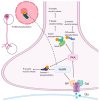
Potential presynaptic signaling cascades activated by AMPAR. AMPARs can activate mitogen-associated protein kinase (MAPK) and protein kinase A (PKA). MAPK phosphorylates synapsin I, an event that leads to an increase in evoked neurotransmitter release by increasing the availability of vesicles for fusion. PKA has many different substrates. Among them, Snapin phosphorylation enhances the association of synaptotagmin with the soluble N-ethylmaleimide-sensitive factor attachment protein receptor (SNARE) complex. PKA increases cysteine string protein (CSP) phosphorylation lowering its affinity for synaptotagmin I and syntaxin, slows vesicular release with a concomitant increase in quantal size. Created in BioRender.com (accessed on 27 August 2021).
Figure 4
Figure 4
Basic mechanisms underlying AMPAR functions at the presynapse. AMPAR activity at the presynapse impinges on several pathways: fluctuations in [Ca2+], changes in membrane excitability, MAPK and PKA cascades, Gi/o-activated pathways, neurotransmitter release modulation. Created in BioRender.com (accessed on 27 August 2021).

References

    1. Torgner I., Kvamme E. Synthesis of Transmitter Glutamate and the Glial-Neuron Interrelationship. Mol. Chem. Neuropathol. 1990;12:11–17. doi: 10.1007/BF03160053. - DOI - PubMed
    1. Farsi Z., Jahn R., Woehler A. Proton Electrochemical Gradient: Driving and Regulating Neurotransmitter Uptake. BioEssays. 2017;39:1600240. doi: 10.1002/bies.201600240. - DOI - PubMed
    1. Niswender C.M., Conn P.J. Metabotropic Glutamate Receptors: Physiology, Pharmacology, and Disease. Annu. Rev. Pharmacol. Toxicol. 2010;50:295–322. doi: 10.1146/annurev.pharmtox.011008.145533. - DOI - PMC - PubMed
    1. Traynelis S.F., Wollmuth L.P., McBain C.J., Menniti F.S., Vance K.M., Ogden K.K., Hansen K.B., Yuan H., Myers S.J., Dingledine R. Glutamate Receptor Ion Channels: Structure, Regulation, and Function. Pharmacol. Rev. 2010;62:405–496. doi: 10.1124/pr.109.002451. - DOI - PMC - PubMed
    1. Möykkynen T., Coleman S.K., Semenov A., Keinänen K. The N-Terminal Domain Modulates α-Amino-3-Hydroxy-5-Methyl-4-Isoxazolepropionic Acid (AMPA) Receptor Desensitization. J. Biol. Chem. 2014;289:13197–13205. doi: 10.1074/jbc.M113.526301. - DOI - PMC - PubMed

Publication types

MeSH terms

Substances