. 2021 Aug 31;10(9):2256.
doi: 10.3390/cells10092256.

Free Transplantation of a Tissue Engineered Bone Graft into an Irradiated, Critical-Size Femoral Defect in Rats

Affiliations

Free Transplantation of a Tissue Engineered Bone Graft into an Irradiated, Critical-Size Femoral Defect in Rats

Ulrike Rottensteiner-Brandl et al. Cells. .

Abstract

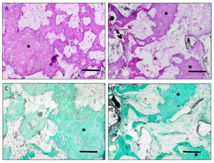
Healing of large bone defects remains a challenge in reconstructive surgery, especially with impaired healing potential due to severe trauma, infection or irradiation. In vivo studies are often performed in healthy animals, which might not accurately reflect the situation in clinical cases. In the present study, we successfully combined a critical-sized femoral defect model with an ionizing radiation protocol in rats. To support bone healing, tissue-engineered constructs were transferred into the defect after ectopic preossification and prevascularization. The combination of SiHA, MSCs and BMP-2 resulted in the significant ectopic formation of bone tissue, which can easily be transferred by means of our custom-made titanium chamber. Implanted osteogenic MSCs survived in vivo for a total of 18 weeks. The use of SiHA alone did not lead to bone formation after ectopic implantation. Analysis of gene expression showed early osteoblast differentiation and a hypoxic and inflammatory environment in implanted constructs. Irradiation led to impaired bone healing, decreased vascularization and lower short-term survival of implanted cells. We conclude that our model is highly valuable for the investigation of bone healing and tissue engineering in pre-damaged tissue and that healing of bone defects can be substantially supported by combining SiHA, MSCs and BMP-2.

Keywords: bone tissue engineering; critical size defect; hydroxyapatite; irradiation; mesenchymal stem cells.


Conflict of interest statement

The authors declare no conflict of interest.

Figures

Figure 1
Figure 1
Implantation of chambers into donor animals. The chamber was filled with hydroxyapatite scaffold and fibrin sealant with or without rhBMP-2/mesenchymal stem cells after osteogenic differentiation (A,B). Afterward, the chamber was secured to the muscles in the medial femoral region (C,D).
Figure 2
Figure 2
CSFD surgery. After exposure of the femur (A), Kirschner wire pins were positioned using an empty template chamber (BD), and a 10 mm piece of the femoral shaft was removed (E). The prevascularized construct was transferred from the donor to the acceptor animal and secured with screws and a cerclage wire (FH).
Figure 3
Figure 3
Bone formation in implanted constructs. The newly formed bone tissue was clearly visible in group C/C irrad and D/D irrad (white arrows), whereas no bone formation was induced by the scaffold alone (B/B irrad). Outgrowth of bone tissue from the femoral stumps was present in group A but very limited in group A irrad (black arrows). ((AC): 12 weeks after CSFD, (D): 10 days after CSFD). Scale bar 2 mm. Merged images. Total magnification ×40.
Figure 4
Figure 4
Relative bone area in constructs after 12 weeks (group A/A irrad, B/B irrad, C/C irrad) and 10 days (group D/D irrad). Bone formation was significantly impaired in group A irrad, compared to group A (* p ≤ 0.05). No bone formation was noted in constructs filled with hydroxyapatite scaffold alone (B/B irrad). Compared to the short-term groups (D/D irrad), the relative bone area increased in unirradiated defects (group C), whereas cessation of ossification occurred in irradiated bones (C irrad). This led to a significantly higher relative bone area in group C compared to group C irrad (* p ≤ 0.05). The implantation of hydroxyapatite/BMP-2/MSCs led to a significantly higher ossification than in empty defects or defects filled with hydroxyapatite alone, both with and without irradiation (* p ≤ 0.05; ** p ≤ 0.01, *** p ≤ 0.001). Values are given as mean ± SD.
Figure 5
Figure 5
Representative immunohistochemical staining of constructs of group C (A,C,E) and C irrad (B,D,F). ((A,B): Lectin staining; (C,D): alpha-smooth muscle actin staining; (E,F): CD68 staining). (AD): Vessel lumina filled with Microfil® (black dots, red arrows) were lectin and alpha-smooth muscle actin positive in groups C and C irrad. (E,F): In groups C and C irrad, there were CD68 positive cells around the granule detectable. (Arrows: Microfil® inside the vessel lumen; Asterisk: degraded scaffold). Scale bar 0.2 mm. Total magnification ×40.
Figure 6
Figure 6
H&E and Masson Goldners’ trichrome staining of bone formations in group C and C irrad (Asterisk: bone formations; (A,C): group C; (B,D): group C irrad, (A,B): H&E staining; (C,D): Masson Goldners’ trichrome staining). Scale bar 0.2 mm. Total magnification ×40.
Figure 7
Figure 7
Masson Goldners’ trichrome staining of bone formations. The bone generation was detectable in group C/C irrad and D/D irrad, whereas no bone formation was induced by the scaffold alone (B/B irrad). Outgrowth of bone tissue from the femoral stumps was present in group A but very limited in group A irrad (black arrows). ((AC): 12 weeks after CSFD, (D): 10 days after CSFD). Scale bar 2 mm. Merged images. Total magnification ×40.
Figure 8
Figure 8
Vascularization of implanted constructs 12 weeks (group A/B/C, A irrad/B irrad/C irrad) and 10 days (D/D irrad) after CSFD surgery. (A) The relative vessel area was low in short-term groups D and D irrad and increased after 12 weeks in all groups. Empty defects (group A) showed a significantly higher relative vessel area than defects filled with hydroxyapatite alone (group B; ** p ≤ 0.01) or hydroxyapatite/BMP-2/MSCs (group C; * p ≤ 0.05). The differences between irradiated defects and unirradiated controls were not significant. (B) Similar to the relative vessel area, vessels in empty defects (group A) were significantly larger than hydroxyapatite-filled defects (group B; * p ≤ 0.05). The differences between irradiated and unirradiated groups were not significant. Values are given as mean ± SD.
Figure 9
Figure 9
Survival of implanted, DiI labeled osteogenic MSCs in constructs, 12 weeks (group C/C irrad) and 10 days (D/D irrad) after CSFD surgery. (A) A significantly lower cell survival was noted in irradiated femora 10 days after CSFD, compared to unirradiated controls (** p ≤ 0.01). After 12 weeks, the difference was not significant. (B) In all groups, a significantly higher number of cells survived in the periphery compared to the center of constructs (*** p ≤ 0.001). However, the spatial distribution did not differ between different groups. Values are given as mean ± SD. (CF): Representative images of DiI positive cells (red) and DAPI staining (blue) ((C): group C; (D): group C irrad, (E): group D; (F): group D irrad) (Total magnification ×100, Scale bar 50 µm).
Figure 10
Figure 10
Gene expression of osteogenic, fibrotic, hypoxic, angiogenic and inflammatory markers in irradiated and unirradiated constructs, compared to healthy bone tissue (bone tissue set 1). A significant up or downregulation in comparison to bone tissue is marked with * (p ≤ 0.05). A significant difference in osteocalcin expression between group C and C irrad was noted (§, p ≤ 0.05). Values are given as mean ± SD.

References

    1. Toogood P., Miclau T. Critical-Sized Bone Defects: Sequence and Planning. J. Orthop. Trauma. 2017;31:S23–S26. doi: 10.1097/BOT.0000000000000980. - DOI - PMC - PubMed
    1. Landau M.J., Badash I., Yin C., Alluri R.K., Patel K.M. Free vascularized fibula grafting in the operative treatment of malignant bone tumors of the upper extremity: A systematic review of outcomes and complications. J. Surg. Oncol. 2018;117:1432–1439. doi: 10.1002/jso.25032. - DOI - PubMed
    1. Niu Y., Bai Y., Xu S., Wu D., Liu X., Wang P., Zhang C., Li M. Treatment of bone nonunion and bone defects associated with unsuccessful humeral condylar fracture repair with autogenous iliac bone reconstruction. J. Shoulder Elb. Surg. 2012;21:985–991. doi: 10.1016/j.jse.2011.06.004. - DOI - PubMed
    1. Qiu X.-S., Chen Y.-X., Qi X.-Y., Shi H.-F., Wang J.-F., Xiong J. Outcomes of cement beads and cement spacers in the treatment of bone defects associated with post-traumatic osteomyelitis. BMC Musculoskelet. Disord. 2017;18:256. doi: 10.1186/s12891-017-1614-1. - DOI - PMC - PubMed
    1. García-Gareta E., Coathup M.J., Blunn G.W. Osteoinduction of bone grafting materials for bone repair and regeneration. Bone. 2015;81:112–121. doi: 10.1016/j.bone.2015.07.007. - DOI - PubMed

Publication types