. 2021 Sep 5;10(9):2322.
doi: 10.3390/cells10092322.

Biosynthesis of the Novel Endogenous 15-Lipoxygenase Metabolites N-13-Hydroxy-octodecadienoyl-ethanolamine and 13-Hydroxy-octodecadienoyl-glycerol by Human Neutrophils and Eosinophils

Affiliations

Biosynthesis of the Novel Endogenous 15-Lipoxygenase Metabolites N-13-Hydroxy-octodecadienoyl-ethanolamine and 13-Hydroxy-octodecadienoyl-glycerol by Human Neutrophils and Eosinophils

Anne-Sophie Archambault et al. Cells. .

Abstract

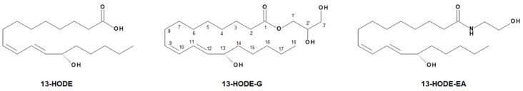
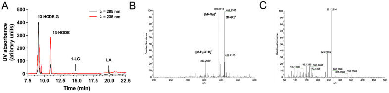
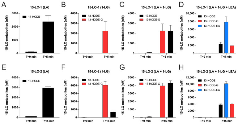
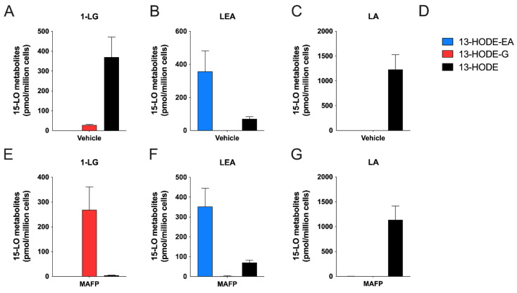
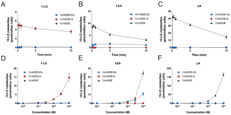
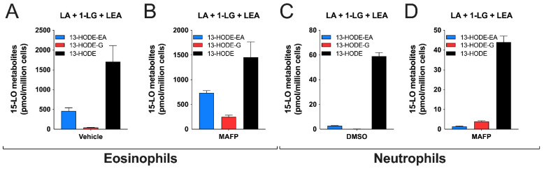
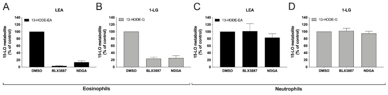
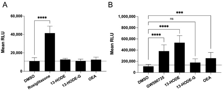
The endocannabinoids 2-arachidonoyl-glycerol and N-arachidonoyl-ethanolamine are lipids regulating many physiological processes, notably inflammation. Endocannabinoid hydrolysis inhibitors are now being investigated as potential anti-inflammatory agents. In addition to 2-arachidonoyl-glycerol and N-arachidonoyl-ethanolamine, the endocannabinoidome also includes other monoacylglycerols and N-acyl-ethanolamines such as 1-linoleoyl-glycerol (1-LG) and N-linoleoyl-ethanolamine (LEA). By increasing monoacylglycerols and/or N-acyl-ethanolamine levels, endocannabinoid hydrolysis inhibitors will likely increase the levels of their metabolites. Herein, we investigated whether 1-LG and LEA were substrates for the 15-lipoxygenase pathway, given that both possess a 1Z,4Z-pentadiene motif, near their omega end. We thus assessed how human eosinophils and neutrophils biosynthesized the 15-lipoxygenase metabolites of 1-LG and LEA. Linoleic acid (LA), a well-documented substrate of 15-lipoxygenases, was used as positive control. N-13-hydroxy-octodecadienoyl-ethanolamine (13-HODE-EA) and 13-hydroxy-octodecadienoyl-glycerol (13-HODE-G), the 15-lipoxygenase metabolites of LEA and 1-LG, were synthesized using Novozym 435 and soybean lipoxygenase. Eosinophils, which express the 15-lipoxygenase-1, metabolized LA, 1-LG, and LEA into their 13-hydroxy derivatives. This was almost complete after five minutes. Substrate preference of eosinophils was LA > LEA > 1-LG in presence of 13-HODE-G hydrolysis inhibition with methyl-arachidonoyl-fluorophosphonate. Human neutrophils also metabolized LA, 1-LG, and LEA into their 13-hydroxy derivatives. This was maximal after 15-30 s. Substrate preference was LA ≫ 1-LG > LEA. Importantly, 13-HODE-G was found in humans and mouse tissue samples. In conclusion, our data show that human eosinophils and neutrophils metabolize 1-LG and LEA into the novel endogenous 15-lipoxygenase metabolites 13-HODE-G and 13-HODE-EA. The full biological importance of 13-HODE-G and 13-HODE-EA remains to be explored.

Keywords: 13-HODE; 2-arachidonoyl-glycerol; N-linoleoyl-ethanolamine; anandamide; eicosanoid; endocannabinoid; eosinophils; linoleic acid; linoleoyl-glycerol; neutrophils.


Conflict of interest statement

The authors declare no conflict of interest. The funders had no role in the design of the study; in the collection, analyses, or interpretation of data; in the writing of the manuscript, or in the decision to publish the results.

Figures

Figure 1
Figure 1
Chemical structures of 13-HODE, 13-HODE-G, and 13-HODE-EA.
Figure 2
Figure 2
RP-HPLC chromatogram and mass spectrometry analysis of 13-HODE-G. (A) Separation of 13-HODE-G from other possible contaminants. The reaction products (~350 µg) of 1-LG with soybean lipoxygenase were loaded onto the HPLC column and eluted as described in Material and Methods. (B) Positive electrospray ionization mass spectrometry (ESI+) of 13-HODE-G. Positive electrospray ionization mass spectrometry (ESI+) of 13-HODE-G yielded three main cations; [M-H2O+H]+ was found at m/z 353.2689, [M+Na]+ at m/z 393.2618 and [M+K]+ at m/z 409.2355. (C) Collision-Induced Dissociation (CID) at Higher-energy collisional dissociation (HCD) of 20 eV of 13-HODE-G. CID of [M-H2O+H]+ at m/z 353.2689 led to the dominant product ion of m/z 261.2214.
Figure 3
Figure 3
Metabolism of 1-LG, LA and LEA by human recombinant 15-LO-1 and 15-LO-2. Human recombinant 15-LO-1 (AD) or 15-LO-2 (EH) were incubated at 37 °C in HEPES 25 mM containing 0.01% triton. A total of 10 μM of LA (A,D,E,H); 1-LG (B,D,F,H) or LEA (D,H) were added for 5 min (15-LO-1) or 15 min (15-LO-2). NaBH4 was then added for 5 min and incubations were stopped by the addition of 0.5 mL of cold (−20 °C) MeOH containing 0.01% acetic acid and the internal standards. Samples then were processed for LC-MS/MS analyses as described in methods. Results are the mean (±SEM) of 3 independent experiments.
Figure 4
Figure 4
Metabolism of LA, 1-LG or LEA by human eosinophils. Pre-warmed human eosinophil suspensions (37 °C, 106 cells/mL in HBSS containing 1.6 mM CaCl2) were treated with DMSO (AC) or MAFP 1 μM (EG) for 5 min then stimulated with 3 μM of (A,E) 1-LG, (B,F) LEA, (C,G) LA for another 5 min. (D) Metabolite legend for panels. (AG) Incubations were stopped by the addition of one volume of cold (−20 °C) MeOH containing the internal standards. Samples then were processed for LC-MS analysis as described in methods. Results are the mean (±SEM) of 4–5 independent experiments.
Figure 5
Figure 5
Time- and concentration-dependent synthesis of 15-LO metabolites from 1-LG, LEA, and LA by human eosinophils. Pre-warmed human eosinophils suspensions (106 cells/mL in HBSS containing 1.6 mM CaCl2) were treated with 3 μM of (A) 1-LG, (B) LEA, or (C) LA for the indicated times. (DF) Eosinophils were treated with increasing concentration of (D) 1-LG, (E) LEA or (F) LA for 5 min. In experiments involving 1-LG, cells were pretreated with MAFP for 5 min prior to the addition of 1-LG. Incubations were stopped by adding one volume of cold (−20 °C) MeOH containing the internal standards. Samples then were processed for LC-MS/MS analyses as described in methods. Data are the mean (±SEM) of 4–5 independent experiments.
Figure 6
Figure 6
15-LO metabolite production in response to LA, 1-LG, or LEA by human neutrophils. Pre-warmed human neutrophil suspensions (37 °C, 5 × 10⁶ cells/mL in HBSS containing 1.6 mM CaCl2) were pre-treated with DMSO (AC) or MAFP (EG) for 5 min before the addition of 3 μM of either (A,E) 1-LG, (B,F) LEA, (C,G) LA. (D) Metabolite legend for panels AG. Incubations were stopped after 5 min by the addition of one volume of cold (−20 °C) MeOH containing the internal standards. Samples then were processed for LC-MS/MS analyses as described in methods. Results are the mean (±SEM) of 4 independent experiments.
Figure 7
Figure 7
Time- and concentration-dependent biosynthesis of 15-LO metabolites from 1-LG, LEA, and LA by human neutrophils. Pre-warmed human neutrophils suspensions (37 °C, 5 × 10⁶ cells/mL in HBSS containing 1.6 mM CaCl2) were treated with 3 μM of (A) 1-LG, (B) LEA, or (C) LA for the indicated times. (DF) Neutrophils were treated with increasing concentration of (D) 1-LG, (E) LEA, or (F) LA for 5 min. In experiments involving 1-LG, cells were pretreated with 1 µM MAFP for 5 min prior to the addition of 1-LG. Incubations were stopped by the addition of one volume of cold (−20 °C) MeOH containing the internal standards. Samples then were processed for LC-MS/MS analyses as described in methods. Data are the mean (±SEM) of 4 independent experiments.
Figure 8
Figure 8
Substrate preference by human eosinophils and neutrophils. Pre-warmed (37 °C) human eosinophil suspensions (106 cells/mL in HBSS containing 1.6 mM CaCl2) or neutrophil suspensions (5 × 10⁶ cells/mL in HBSS containing 1.6 mM CaCl2) were pre-treated with DMSO (A,C) or MAFP (B,D) for 5 min before the addition of a mixture of 1-LG, LEA, and LA at 3 μM each. Incubations were stopped after 5 min by the addition of one volume of cold (−20 °C) MeOH containing the internal standards. Samples then were processed for LC-MS/MS analyses as described in methods. Results are the mean (±SEM) of 4 independent experiments.
Figure 9
Figure 9
Inhibition of 13-HODE-G and 13-HODE-EA biosynthesis by 15-LO inhibitors in human eosinophils and neutrophils. Pre-warmed (37 °C) human (A,B) eosinophil suspensions (106 cells/mL in HBSS containing 1.6 mM CaCl2) or (C,D) neutrophil suspensions (5 × 10⁶ cells/mL in HBSS containing 1.6 mM CaCl2) were pre-treated with DMSO, BLX-3887 (10 µM) or NDGA (10 µM) for 5 min before the addition of 3 μM (A,C) LEA or (B,D) 1-LG. Incubations were stopped 5 min after LEA or 1-LG treatment by the addition of one volume of cold (−20 °C) MeOH containing the internal standards. Samples then were processed for LC-MS/MS analyses as described in methods. Results are the mean (±SEM) of 4 independent experiments.
Figure 10
Figure 10
Impact of 13-HODE and 13-HODE-G on PPAR activity. Cells were incubated with 30 µM 13-HODE, 13-HODE-G, or OEA overnight and PPAR activity was measured using the PPAR assay kits according to the manufacturers’ instructions. Rosiglitazone (10 µM) and GW 590735 (100 nM) were utilized as positive controls for the (A) PPAR-γ and (B) PPAR-α assays, respectively. Data represent the mean (± SEM) of three (PPAR-γ) or five (PPAR-α) experiments, each performed in triplicates. p values were obtained by performing a 2-way ANOVA with a Sidak multiple comparison tests. **** p < 0.0001, *** p < 0.001. ns: p > 0.05.
Figure 11
Figure 11
Representative frames from molecular dynamics of PPARα (light gray) in complex with 13-HODE (dark grey) shown in ball and stick representation. Panel (A,B) show the docking pose I and pose II of the ligand, respectively. Protein residues within 5 Å from the ligands are shown in stick representation. H-bonds are shown as green stick. Hydrogen, nitrogen, oxygen, and sulfur atom are painted white, blue, red, and yellow, respectively. A transparent surface for ribbons was used wherever they hide the ligand-binding site.

References

    1. Turcotte C., Chouinard F., Lefebvre J.S., Flamand N. Regulation of inflammation by cannabinoids, the endocannabinoids 2-arachidonoyl-glycerol and arachidonoyl-ethanolamide, and their metabolites. J. Leukoc. Biol. 2015;97:1049–1070. doi: 10.1189/jlb.3RU0115-021R. - DOI - PubMed
    1. Di Marzo V. New approaches and challenges to targeting the endocannabinoid system. Nat. Rev. Drug Discov. 2018;17:623–639. doi: 10.1038/nrd.2018.115. - DOI - PubMed
    1. Prescott S.M., Majerus P.W. Characterization of 1,2-diacylglycerol hydrolysis in human platelets. Demonstration of an arachidonoyl-monoacylglycerol intermediate. J. Biol. Chem. 1983;258:764–769. doi: 10.1016/S0021-9258(18)33114-4. - DOI - PubMed
    1. Goparaju S.K., Ueda N., Taniguchi K., Yamamoto S. Enzymes of porcine brain hydrolyzing 2-arachidonoylglycerol, an endogenous ligand of cannabinoid receptors. Biochem. Pharmacol. 1999;57:417–423. doi: 10.1016/S0006-2952(98)00314-1. - DOI - PubMed
    1. Xie S., Borazjani A., Hatfield M.J., Edwards C.C., Potter P.M., Ross M.K. Inactivation of lipid glyceryl ester metabolism in human THP1 monocytes/macrophages by activated organophosphorus insecticides: Role of carboxylesterases 1 and 2. Chem. Res. Toxicol. 2010;23:1890–1904. doi: 10.1021/tx1002194. - DOI - PMC - PubMed

Publication types

LinkOut - more resources