. 2021 Sep 6;10(9):2336.
doi: 10.3390/cells10092336.

Identification of GOLPH3 Partners in Drosophila Unveils Potential Novel Roles in Tumorigenesis and Neural Disorders

Affiliations

Identification of GOLPH3 Partners in Drosophila Unveils Potential Novel Roles in Tumorigenesis and Neural Disorders

Stefano Sechi et al. Cells. .

Abstract

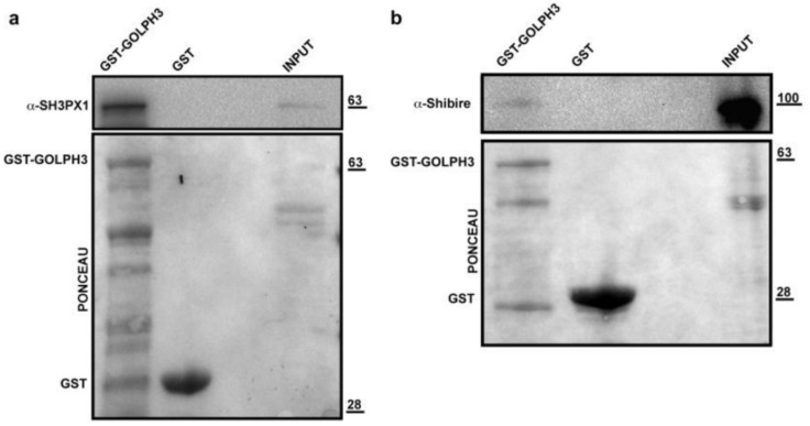
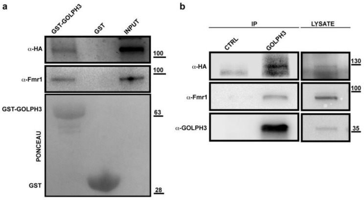
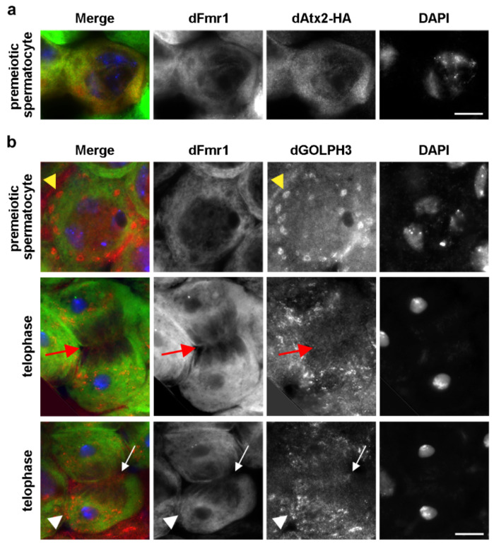
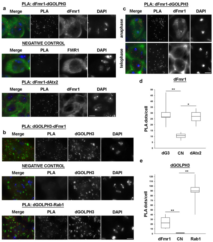
Golgi phosphoprotein 3 (GOLPH3) is a highly conserved peripheral membrane protein localized to the Golgi apparatus and the cytosol. GOLPH3 binding to Golgi membranes depends on phosphatidylinositol 4-phosphate [PI(4)P] and regulates Golgi architecture and vesicle trafficking. GOLPH3 overexpression has been correlated with poor prognosis in several cancers, but the molecular mechanisms that link GOLPH3 to malignant transformation are poorly understood. We recently showed that PI(4)P-GOLPH3 couples membrane trafficking with contractile ring assembly during cytokinesis in dividing Drosophila spermatocytes. Here, we use affinity purification coupled with mass spectrometry (AP-MS) to identify the protein-protein interaction network (interactome) of Drosophila GOLPH3 in testes. Analysis of the GOLPH3 interactome revealed enrichment for proteins involved in vesicle-mediated trafficking, cell proliferation and cytoskeleton dynamics. In particular, we found that dGOLPH3 interacts with the Drosophila orthologs of Fragile X mental retardation protein and Ataxin-2, suggesting a potential role in the pathophysiology of disorders of the nervous system. Our findings suggest novel molecular targets associated with GOLPH3 that might be relevant for therapeutic intervention in cancers and other human diseases.

Keywords: Drosophila; FMRP; GOLPH3; Golgi; cell cycle; male meiosis; spermatogenesis.


Conflict of interest statement

The authors declare no competing or financial interests.

Figures

Figure 1
Figure 1
dGOLPH3-RFP localizes to the multiple Golgi organelles in primary spermatocytes and to the acroblast in spermatids. Fluorescence micrographs of live squashed spermatocytes and spermatids expressing dGOLPH3-RFP and GFP-Cog7. Arrowheads point to Golgi stacks; arrows point to the acroblast. Bar, 10 μm.
Figure 2
Figure 2
Protein Classes and GO analyses. (a) Funnel graph showing the classes of proteins identified in our AP-MS experiments and classified according to PANTHER database [41]. The percentage for each class is indicated. See also Supplementary Table S2 for details. (b) Heat maps showing the GO annotation enrichment profiles of the dGOLPH3 interactome. GO enrichment profiles were analyzed using GOrilla tool [42] under the category “process” and PANTHER database [41] under the category “GO-slim biological process”. Over-represented/enriched GO terms are shown in different color shades according to their fold enrichment as indicated in the color scale bar at the bottom; actual fold enrichment values are shown within the heat map (see Supplementary Tables S3 and S4 for p-values). For simplicity and to improve visual representation, for the PANTHER over-representation analysis only the headings for each GO-slim biological process are shown in the graph, while the full results are reported in the Supplementary Table S4. Only results for Bonferroni-corrected analysis (p < 0.05) were considered.
Figure 3
Figure 3
dGOLPH3 interacts with COPI subunits and Sec31 protein. (a,b) dGOLPH3 protein coprecipitated with RFP–βCOP but not with γCOP-RFP and RFP. Protein extracts from testes expressing RFP–βCOP, γCOP-RFP (a) and RFP (RFP IP CTRL) (b), were immunoprecipitated with RFP-trap beads (α-RFP) and blotted for either RFP or dGOLPH3. 4% of the total lysates and one third of the IP were loaded and probed with the indicated antibodies. (ce) GST pull-down to test dGOLPH3 interaction with αCOP (c), δCOP (d) and Sec31 (e) proteins. (c) Bacterially expressed GST and GST-dGOLPH3 were purified by Gluthatione-Sepharose beads, incubated with testis protein extracts from Oregon-R males and blotted for αCOP protein. (d) Bacterially expressed GST and GST-GOLPH3 were purified by Gluthatione-Sepharose beads, incubated with testis protein extracts from males expressing δCOP-HA. (e) Bacterially expressed GST-dGOLPH3 and GST purified by Gluthatione-Sepharose beads were incubated with testis protein extracts from Oregon-R males and blotted for Sec31. Ponceau staining in (ce) is shown as a loading control. 2% of the input and 25% of the pull-downs were loaded and probed with the indicated antibody. Molecular masses in (ae), expressed in kilodaltons.
Figure 4
Figure 4
dGOLPH3 interacts with vesicle trafficking and cytoskeleton proteins. (a) Protein extracts from testes expressing either GFP or the indicated YFP-tagged Rab protein were immunoprecipitated with GFP traps and blotted to detect either GFP/YFP or GOLPH3. 4% of the total lysates and one third of IP were loaded and probed with the indicated antibodies. (b) Protein extracts from Drosophila testes expressing GFP (control) and GFP tagged Rac1, Rac2 and Sec22 were immunoprecipitated with GFP trap and blotted to detects either GFP or GOLPH3. 4% of the total lysates and one third of IP were loaded and probed with the indicated antibodies. Molecular masses in (a,b) are expressed in kilodaltons.
Figure 5
Figure 5
dGOLPH3 interacts with endocytic trafficking proteins. GST (control) and recombinant GST-GOLPH3 proteins, immobilized on Gluthatione-Sepharose beads were incubated with testis protein extracts from Oregon-R males. Ponceau staining is shown as a loading control. 2% of the input and 25% of the pull-down were loaded and probed for SH3PX1 (a) and Shibire (b). Molecular masses are expressed in kilodaltons.
Figure 6
Figure 6
dGOLPH3 interacts with dAtx2 and dFmr1. (a) Bacterially expressed GST-dGOLPH3 and GST (control) were purified by Gluthatione-Sepharose beads and incubated with testis extracts expressing dAtx2-HA. Ponceau staining is shown as a loading control. A percentage of 2% of the input and 25% of pull-downs were loaded and probed with the indicated antibody. Molecular mass is expressed in kilodaltons. (b) Protein extracts from testes expressing dAtx2-HA were immunoprecipitated with antibodies against Drosophila GOLPH3 (rabbit G49139/77) and blotted with mouse anti-dGOLPH3 S11047/1/56, mouse anti-dFmr1, or mouse anti-HA. Pre-immune serum (G49139/1, from the same animal before the immunization) was used in control experiments (CTRL). Molecular mass is expressed in kilodaltons.
Figure 7
Figure 7
dFmr1 localization in premeiotic and dividing spermatocytes. (a) dFmr1 co-localizes with dAtx2 in the cytoplasm of primary spermatocytes. Testes expressing dAtx2-HA were stained for dFmr1(green), HA (dAtx2, red) and DNA (DAPI, blue). n = 40 spermatocytes randomly selected from images taken in five independent experiments. (b) Premeiotic and dividing spermatocytes were stained for dFmr1 (green) dGOLPH3 (red) and DNA (DAPI, blue). dFmr1 localizes to the cytoplasm of premeiotic spermatocytes and concentrates at the midzone (arrows) and at the astral membranes (arrowheads) at each pole of dividing spermatocytes (telophase). dGOLPH3 protein is visible in the cytoplasm and enriched in the Golgi stacks of premeiotic spermatocytes (yellow arrowheads). During telophase dGOLPH3 protein localizes to the midzone (arrows) and is enriched in puncta at the astral membranes of dividing spermatocytes (arrowheads). n = 40 premeiotic spermatocytes and n = 25 telophase spermatocytes, randomly selected from images taken in five independent experiments. Bars, 10 μm.
Figure 8
Figure 8
dFmr1 interacts with dAtx2 and dGOLPH3 in primary spermatocytes. (a,b) Proximity ligation assay (PLA) to visualize dFmr1-dGOLPH3 and dFmr1-dAtx2 interactions in premeiotic spermatocytes. In negative control primary anti-GOLPH3 (a) or anti-dFmr1 (b) antibodies were omitted. dGOLPH3-Rab1 interaction was used in positive control experiments. (c) PLA to visualize dFmr1-dGOLPH3 interaction in dividing spermatocytes. (d,e) Quantification of the PLA signals per cell was obtained as described in Materials and Methods. The box plots show the number of PLA dots per cell in dFmr1-dGOLPH3, dFmr1-dAtx2 and dGOLPH3-Rab1compared to negative controls (CN). dG3, dGOLPH3. A total of 50 cells were randomly selected from images taken in 5 independent experiments. Statistic significant differences are * p < 0.05; ** p < 0.01 (Mann-Whitney test). Bars, 10 μm.

Similar articles

Cited by

References

    1. Dippold H.C., Ng M.M., Farber-Katz S.E., Lee S.K., Kerr M.L., Peterman M.C., Sim R., Wiharto P.A., Galbraith K.A., Madhavarapu S., et al. GOLPH3 bridges phosphatidylinositol-4- phosphate and actomyosin to stretch and shape the Golgi to promote budding. Cell. 2009;139:337–351. doi: 10.1016/j.cell.2009.07.052. - DOI - PMC - PubMed
    1. Iyer P., Bhave M., Jain B.K., RoyChowdhury S., Bhattacharyya D. Vps74p controls Golgi size in an Arf1-dependent manner. FEBS Lett. 2018;592:3720–3735. doi: 10.1002/1873-3468.13266. - DOI - PubMed
    1. Rahajeng J., Kuna R.S., Makowski S.L., Tran T.T.T., Buschman M.D., Li S., Cheng N., Ng M.M., Field S.J. Efficient Golgi Forward Trafficking Requires GOLPH3-Driven, PI4P-Dependent Membrane Curvature. Dev. Cell. 2019;50:573–585. doi: 10.1016/j.devcel.2019.05.038. - DOI - PMC - PubMed
    1. Sechi S., Colotti G., Belloni G., Mattei V., Frappaolo A., Giansanti M.G. GOLPH3 Is Essential for Contractile Ring Formation and Rab11 Localization to the Cleavage Site during Cytokinesis in Drosophila melanogaster. PLoS Genet. 2014;10:e1004305. doi: 10.1371/journal.pgen.1004305. - DOI - PMC - PubMed
    1. Sechi S., Frappaolo A., Karimpour-Ghahnavieh A., Piergentili R., Giansanti M.G. Oncogenic Roles of GOLPH3 in the Physiopathology of Cancer. Int. J. Mol. Sci. 2020;21:933. doi: 10.3390/ijms21030933. - DOI - PMC - PubMed

Publication types

MeSH terms

LinkOut - more resources