. 2021 Sep 9;10(9):2370.
doi: 10.3390/cells10092370.

Sex-Dependent Effects of Intestinal Microbiome Manipulation in a Mouse Model of Alzheimer's Disease

Affiliations

Sex-Dependent Effects of Intestinal Microbiome Manipulation in a Mouse Model of Alzheimer's Disease

Harpreet Kaur et al. Cells. .

Abstract

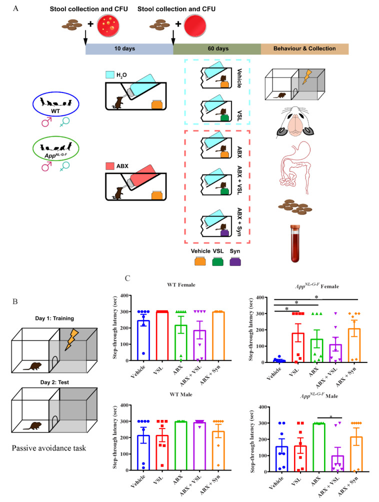
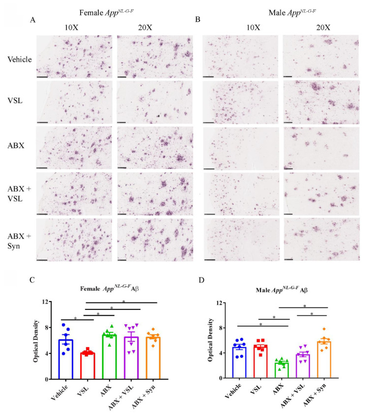
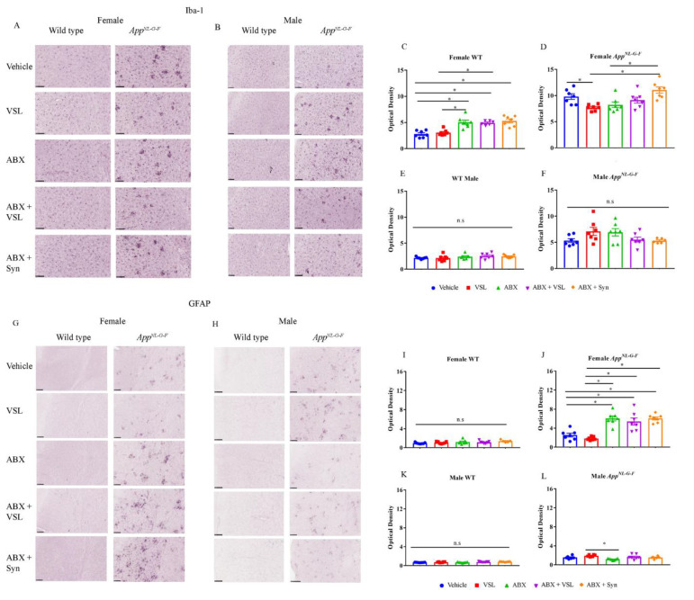
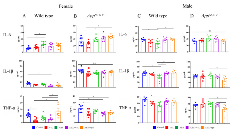
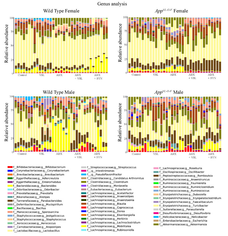
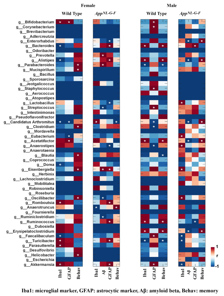
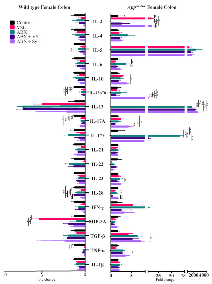
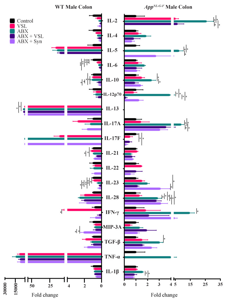
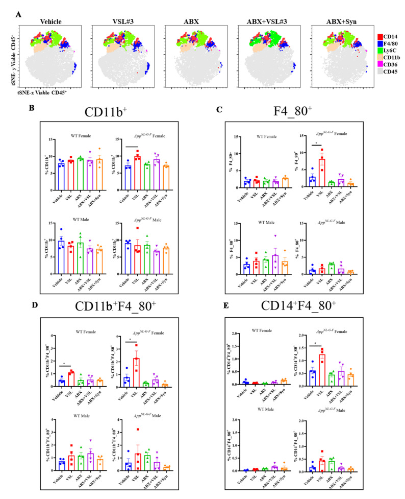
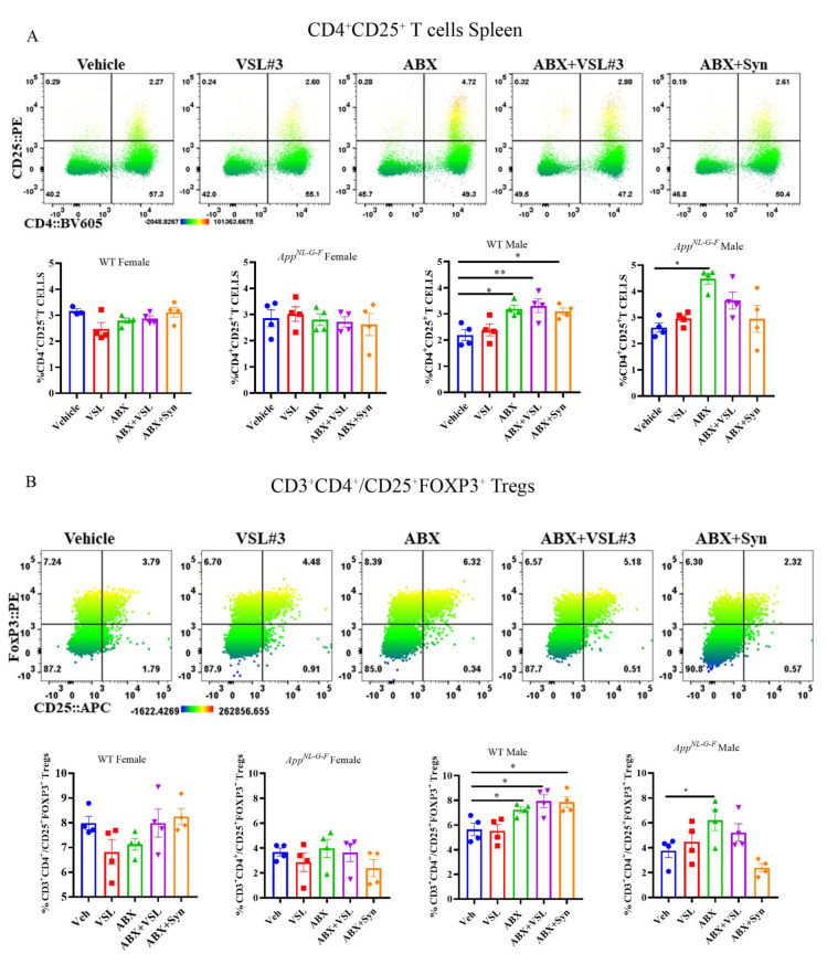
Mechanisms linking intestinal bacteria and neurodegenerative diseases such as Alzheimer's disease (AD) are still unclear. We hypothesized that intestinal dysbiosis might potentiate AD, and manipulating the microbiome to promote intestinal eubiosis and immune homeostasis may improve AD-related brain changes. This study assessed sex differences in the effects of oral probiotic, antibiotics, and synbiotic treatments in the AppNL-G-F mouse model of AD. The fecal microbiome demonstrated significant correlations between bacterial genera in AppNL-G-F mice and Aβ plaque load, gliosis, and memory performance. Female and not male AppNL-G-F mice fed probiotic but not synbiotic exhibited a decrease in Aβ plaques, microgliosis, brain TNF-α, and memory improvement compared to no treatment controls. Although antibiotics treatment did not produce these multiple changes in brain cytokines, memory, or gliosis, it did decrease Aβ plaque load and colon cytokines in AppNL-G-F males. The intestinal cytokine milieu and splenocyte phenotype of female but not male AppNL-G-F mice indicated a modest proinflammatory innate response following probiotic treatment compared to controls, with an adaptive response following antibiotics treatment in male AppNL-G-F mice. Overall, these results demonstrate the beneficial effects of probiotic only in AppNL-G-F females, with minimal benefits of antibiotics or synbiotic feeding in male or female mice.

Keywords: Alzheimer’s disease; VSL#3; antibiotics; cytokines; immune response; microbiome; prebiotic; probiotic; splenocytes; synbiotic.


Conflict of interest statement

The authors declare no conflict of interest.

Figures

Figure 1
Figure 1
Study design and treatment effects of probiotic and antibiotics on learning and memory. (A) General experimental procedure and a timeline of the study. (B) Animals at 4–5 months of age were assessed for learning and memory using a passive avoidance task. The image shows the neurobehavior training setup, where a mild foot shock was used as an unconditioned stimulus on day 1 (training day) and the step-through latencies were measured when animals entered from a lit compartment to a dark compartment. (C) The passive avoidance task results are shown as step-through latency on day two and are expressed as mean ± SEM. Significant differences were determined by one-way analysis of variance, * p < 0.05 (n = 7).
Figure 2
Figure 2
Effect of probiotic and antibiotics treatments on Aβ accumulation in AppNL-G-F brains. Representative immunohistochemical staining images for Aβ from temporal cortices of mice treated with vehicle, VSL#3, antibiotics (ABX), antibiotics+VSL#3 (ABX + VSL), and antibiotics+VSL#3+prebiotic (ABX + Syn) are shown for (A) female and (B) male AppNL-G-F mice. (C,D) Quantitation of immunostaining was performed from two sections from each animal, and optical density values were averaged and presented as mean ± SEM. Significant differences were determined by a one-way analysis of variance, * p < 0.05 (n = 7). Scale bars are 100 µm (10×) and 50 µm (20×).
Figure 3
Figure 3
Effect of probiotic and antibiotics treatments on Iba1 and GFAP immunoreactivity. Representative immunohistochemical staining images for (A,B) Iba1 and (G,H) GFAP from temporal cortices of female and male WT and AppNL-G-F mice treated with vehicle, VSL#3, antibiotics (ABX), antibiotics+VSL#3 (ABX + VSL), and antibiotics+VSL#3+prebiotic (ABX+Syn) are shown. Quantitation of (CF) Iba1 and (IL) GFAP immunostaining was performed from two sections from each animal, and optical density values were averaged and presented as mean ± SEM. Significant differences were determined by a one-way analysis of variance, * p < 0.05 (n = 7). Scale bars are 50 µm (20×).
Figure 4
Figure 4
Effect of probiotic and antibiotics treatments on proinflammatory cytokines in female and male mice. Protein levels of interleukin-6 (IL-6), IL-1β, and tumor necrosis factor (TNF-α) were quantified from lysates prepared from parietal cortices of WT (A) and AppNL-G-F (B) female mice and WT (C) and AppNL-G-F (D) male mice treated with vehicle, VSL#3, antibiotics (ABX), antibiotics+VSL#3 (ABX + VSL), and antibiotics+VSL#3+prebiotic (ABX + Syn). Lysates were analyzed by enzyme-linked immunosorbent assay (ELISA). Data are presented as mean ± SEM. Significant differences were determined by one-way analysis of variance, * p < 0.05 (n = 7).
Figure 5
Figure 5
Effects of probiotic and antibiotics treatments on gut bacterial diversity in female and male mice. The relative abundance (%) of dominant bacterial genera in fecal samples of C57BL/6 (WT) and AppNL-G-F mice treated with vehicle, VSL#3, antibiotics (ABX), antibiotics+VSL#3 (ABX + VSL), and antibiotics+VSL#3+prebiotic (ABX + Syn) are plotted as bar charts. The relative abundances are based on the proportional frequencies of the DNA sequences classified at the genus level. Seven animals per group were examined. Thus, each color represents a different bacterial genus.
Figure 6
Figure 6
Correlation of Aβ, behavior, and gliosis with fecal microbiome genera in female and male mice. Heatmaps show the Pearson’s correlation between specific gut genera and brain changes observed in control, probiotic-, and antibiotics-treated groups. Blue to red are r values ranging from negative 1 to positive 1: red color, positive correlation; blue color, negative correlation. The asterisk indicates a significant correlation (more than 95%) between genus and brain data.
Figure 7
Figure 7
Quantification of colon levels of Th1, Th2, and Th17 cytokines in female C57BL/6J (WT) and AppNL-G-F mice. Colons were lysed from vehicle, VSL#3, antibiotics (ABX), antibiotics+VSL#3 (ABX + VSL), and antibiotics+VSL#3+prebiotic (ABX + Syn) female WT mice to quantify cytokine levels via commercial slide array. Data are presented as fold change with respect to controls (n = 5). If the controls values were zero, we used 0.1 as an arbitrary value for that group to calculate the fold change in that dataset. Significant differences were determined by a one-way analysis of variance, * p < 0.05, ** p < 0.01.
Figure 8
Figure 8
Quantification of colon levels of Th1, Th2, and Th17 cytokines in male C57BL/6J (WT) and AppNL-G-F mice. Colons were lysed from vehicle, VSL#3, antibiotics (ABX), antibiotics+VSL#3 (ABX+VSL), and antibiotics+VSL#3+prebiotic (ABX + Syn) male WT mice to quantify cytokine levels via commercial slide array. Data are presented as fold change with respect to controls (n = 5). If the controls values were zero, we used 0.1 as an arbitrary value for that group to calculate the fold change in that dataset. Significant differences were determined by a one-way analysis of variance, * p < 0.05, ** p < 0.01.
Figure 9
Figure 9
Upregulation of CD11b+, F4_80+, CD11b+F4_80+, and CD14+F4_80+ macrophages in VSL#3-treated female App NL-G-F mice. Splenocytes were stained with a panel of cell surface markers and measured by flow cytometry. tSNE analysis was run on 4960–5000 live CD45+ single cells per sample using all surface markers, and manually gated populations were overlaid to visualize the subsets. (A) The data presented are representative tSNE plots for female AppNL-G-F mice, generated by concatenating individual samples in each treatment group with the following parameters: iterations: 5000 and perplexity: 30. (BE) Bar graphs show the mean ± SEM of the percentage of CD11b+, F4_80+, CD11b+F4_80+, and CD14+F4_80+ cells across different treatment groups in female and male C57BL/6 (WT) and App NL-G-F mice (n = 3–4 mice/group). Statistically significant differences were computed by a one-way ANOVA, * p < 0.05, ** p < 0.005.
Figure 10
Figure 10
Upregulation of CD4+CD25+ T and FoxP3 expressing CD4+CD25+ T regulatory cells following antibiotics treatment in male WT and App NL-G-F mice. (A) Splenocytes were stained with surface markers directed against a panel of T cell markers and measured by flow cytometry. Representative heat map dot plots from male App NL-G-F mice showing the percentage of CD4+CD25+ T cells gated on viable CD45+CD3+ cells. Numbers in each quadrant indicate the percentage of cells. The heat map color varies from blue to red, indicating relative under and over-representation of the percentage of cells, respectively. Bar graphs display mean ± SEM of the percentage of CD4+CD25+ T cell across different treatment groups in female and male C57BL/6 (WT) and App NL-G-F mice (n = 3–4 mice/group). Statistically significant differences were computed by one-way ANOVA, * p < 0.05, ** p < 0.005. (B) Splenocytes were stained with surface markers directed against a panel of T cell markers and measured by flow cytometry. Representative heat map dot plots from male App NL-G-F mice showing the percentage of CD25+FoxP3+ Tregs gated on viable CD45+/CD3+CD4+ cells. Numbers in each quadrant indicate the percentage of cells. The heat map color varies from blue to red, indicating relative under- and over-representation of the percentage of cells, respectively. Bar graphs display mean ± SEM of percentage of CD25+FoxP3+ Tregs across different treatment groups in female and male C57BL/6 (WT) and App NL-G-F mice (n = 3–4 mice/group). Statistically significant differences were computed by one-way ANOVA, * p < 0.05, ** p < 0.005.

Similar articles

Cited by

References

    1. Clayton T.A., Baker D., Lindon J.C., Everett J.R., Nicholson J.K. Pharmacometabonomic identification of a significant host-microbiome metabolic interaction affecting human drug metabolism. Proc. Natl. Acad. Sci. USA. 2009;106:14728–14733. doi: 10.1073/pnas.0904489106. - DOI - PMC - PubMed
    1. Saha J.R., Butler V.P., Jr., Neu H.C., Lindenbaum J. Digoxin-inactivating bacteria: Identification in human gut flora. Science. 1983;220:325–327. doi: 10.1126/science.6836275. - DOI - PubMed
    1. Dalile B., Van Oudenhove L., Vervliet B., Verbeke K. The role of short-chain fatty acids in microbiota-gut-brain communication. Nat. Rev. Gastroenterol. Hepatol. 2019;16:461–478. doi: 10.1038/s41575-019-0157-3. - DOI - PubMed
    1. Silva Y.P., Bernardi A., Frozza R.L. The Role of Short-Chain Fatty Acids From Gut Microbiota in Gut-Brain Communication. Front. Endocrinol. 2020;11:25. doi: 10.3389/fendo.2020.00025. - DOI - PMC - PubMed
    1. Antonini M., Lo Conte M., Sorini C., Falcone M. How the Interplay Between the Commensal Microbiota, Gut Barrier Integrity, and Mucosal Immunity Regulates Brain Autoimmunity. Front. Endocrinol. 2019;10:1937. doi: 10.3389/fimmu.2019.01937. - DOI - PMC - PubMed

Publication types

MeSH terms

LinkOut - more resources