Review
. 2021 Sep 10;10(9):2383.
doi: 10.3390/cells10092383.

Effects of Extracellular Osteoanabolic Agents on the Endogenous Response of Osteoblastic Cells

Affiliations
Review

Effects of Extracellular Osteoanabolic Agents on the Endogenous Response of Osteoblastic Cells

Giulia Alloisio et al. Cells. .

Abstract

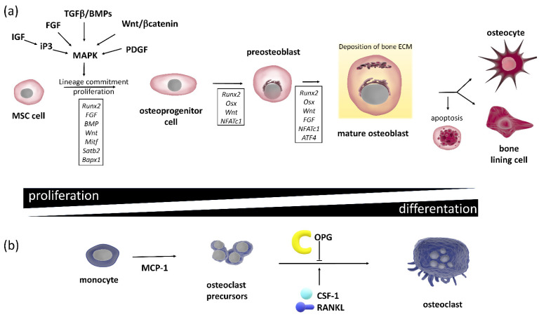
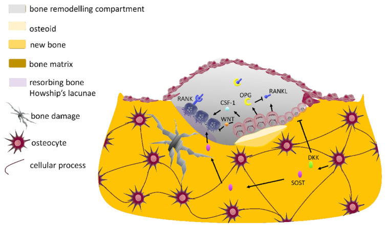
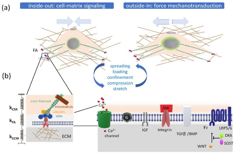
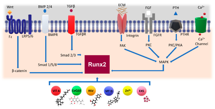
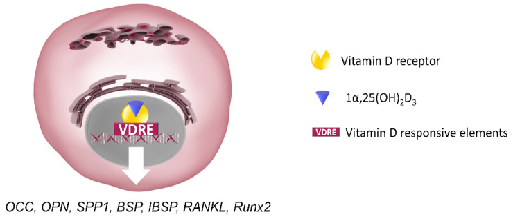
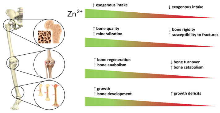
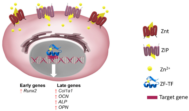
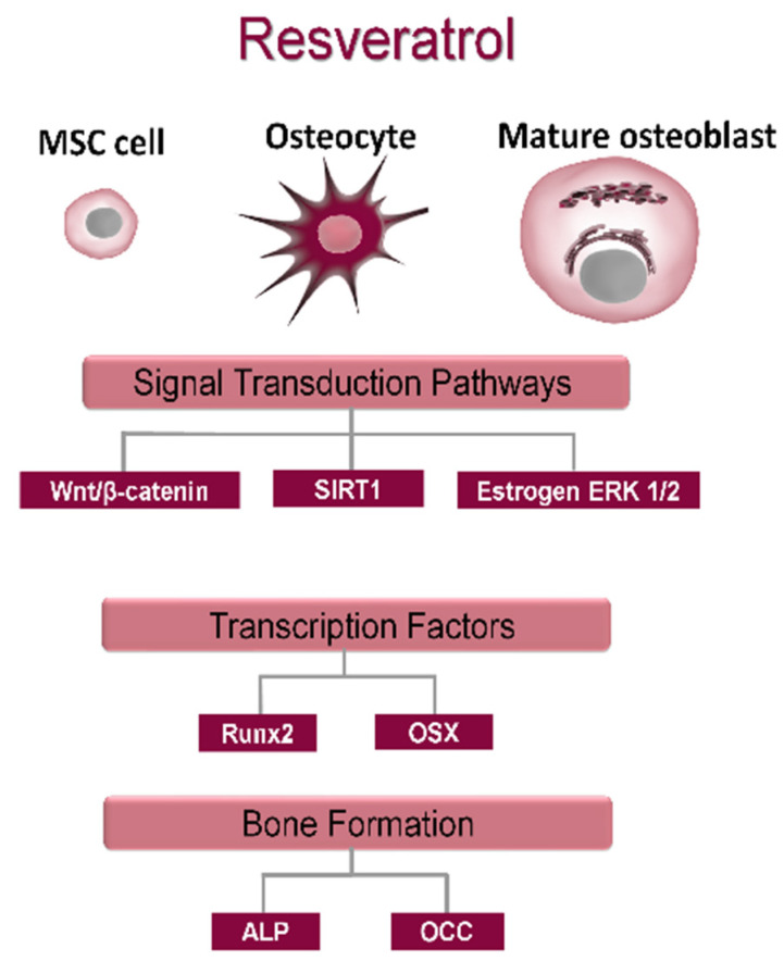
The complex multidimensional skeletal organization can adapt its structure in accordance with external contexts, demonstrating excellent self-renewal capacity. Thus, optimal extracellular environmental properties are critical for bone regeneration and inextricably linked to the mechanical and biological states of bone. It is interesting to note that the microstructure of bone depends not only on genetic determinants (which control the bone remodeling loop through autocrine and paracrine signals) but also, more importantly, on the continuous response of cells to external mechanical cues. In particular, bone cells sense mechanical signals such as shear, tensile, loading and vibration, and once activated, they react by regulating bone anabolism. Although several specific surrounding conditions needed for osteoblast cells to specifically augment bone formation have been empirically discovered, most of the underlying biomechanical cellular processes underneath remain largely unknown. Nevertheless, exogenous stimuli of endogenous osteogenesis can be applied to promote the mineral apposition rate, bone formation, bone mass and bone strength, as well as expediting fracture repair and bone regeneration. The following review summarizes the latest studies related to the proliferation and differentiation of osteoblastic cells, enhanced by mechanical forces or supplemental signaling factors (such as trace metals, nutraceuticals, vitamins and exosomes), providing a thorough overview of the exogenous osteogenic agents which can be exploited to modulate and influence the mechanically induced anabolism of bone. Furthermore, this review aims to discuss the emerging role of extracellular stimuli in skeletal metabolism as well as their potential roles and provide new perspectives for the treatment of bone disorders.

Keywords: antioxidant supplements; bone remodeling; exosomes; mechanically induced anabolism; ossification stimuli; osteoanabolic agents; osteoporosis; resveratrol; retinoic acid; zinc.


Conflict of interest statement

There are no potential conflict of interest relating to the manuscript for each author, and there were no extramural sources supporting this research (excluding sources already declared).

Figures

Figure 1
Figure 1
The principal signaling networks and transcription factors regulating bone cell differentiation. (a) Osteoblastogenesis and osteogenesis. In boxes, transcriptional factors, which characterize each stage of osteogenic differentiation, are shown. The MSC population actively proliferates at the initial stages of osteogenesis. As MSCs commit to osteoblasts, their proliferation rate decreases while they start expressing osteogenic genes. Following mineralization, mature osteoblasts undergo apoptosis, revert back to a bone lining phenotype or become embedded in the mineralized matrix and differentiate into osteocytes. Lines with an arrowhead indicate a positive action, and lines with a bar indicate inhibition. (b) Osteoblast cytokines involved in osteoclastogenesis: osteoblasts produce chemokine MCP-1 (monocyte chemoattractant protein-1). In addition, osteoblasts express the master of osteoclastogenesis cytokines, i.e., CSF-1 (light blue sphere), RANKL (represented in dark blue) and OPG (yellow semicircle). Monocytes (differentiated from HSCs) evolve to osteoclast precursors and finally to active OC forms which are stimulated by RANKL. Together with the canonical Wnt signaling, the RANK/RANKL OPG signaling pathways control osteoclasts in response to the actual extracellular stimuli.
Figure 2
Figure 2
Molecular crosstalk in bone molecular unit (BMU): Paracrine actions of osteoblast-, osteocyte- and osteoclast-derived factors within the bone remodeling compartment. Osteoblasts respond to external signals generated by mechanically activated osteocytes or direct endocrine signals, recruit osteoclast precursors to the remodeling site, by expressing CSF, RANKL (represented in dark blue) and WNT (orange diamond), and inhibit osteoclast activity through OPG (yellow semicircle), a decoy receptor of RANKL (pictured in dark blue). Osteocyte-derived SOST (magenta oval) inhibits OB differentiation and stimulates osteoclastogenesis. The osteocyte expression levels of Wnt inhibitors (SOST and DKK (green oval)) temporally control the cycle of bone remodeling. Lines with an arrowhead indicate a positive action, and lines with a bar indicate inhibition.
Figure 3
Figure 3
(a) Representation of inside-out and outside-in mechanotransduction signals. Focal adhesions (FAs) serve as crucial sites for transferring forces in both directions. Integrins are coupled to the cytoskeleton via molecules such as vinculin, talin and α-actin. (b) Protein network clusters across the extracellular matrix, transmembrane proteins and cytoskeleton regions of a spread cell. On the right side, three nonlinear spring series conceptualize the mechanical linkage between the cytoskeleton, focal adhesion complex and extracellular matrix, with respective nonlinear spring constants: kCSK, kFA, kECM. On the left side, a zoom of the membrane portion is represented. Mechanical signals perceived by membrane-bound receptors such as stretch-activated Ca2+ channels, integrins, G proteins, IGF and TGFβ and/or BMP receptors are stimulated by mechanical forces and converted into a proper biological response (Table 2). The ECM and intracellular pathways are biochemically coupled by mechanotransduction pathways: mechanical resistance to intrinsic forces regulates the stability of focal adhesion complex that contains focal adhesion kinase (FAK), which phosphorylates and activates mechanoresponsive signaling elements. Line with an arrowhead indicates a positive action, and line with a bar indicates inhibition.
Figure 4
Figure 4
The regulation of Runx2 by mechanical and soluble ossification agents. Mechanical signals perceived by membrane-bound receptors such as lipoprotein-related protein (LRP) 5/6 and the frizzled (Fz) co-receptors, TGFβ and/or BMP receptors, integrins, FGF and G proteins (as an example, the PTH receptor is indicated) and stretch-activated Ca2+ channels regulate Rnux2 activity. Major regulatory pathways are represented. Biochemical agents such as vitamin A, coenzyme Q10, resveratrol (RSV), vitamin D, zinc ion and exosomes (EXS) can affect Runx2 and thus osteoblast differentiation.
Figure 5
Figure 5
Scheme of direct actions of 1,25(OH)2D3/VDR on mature osteoblasts. 1,25(OH)2D3 acts via the VDR to regulate the osteoblast-related genes containing VDRE binding motifs. 1,25(OH)2D3 can both positively and negatively regulate expression of osteoblast phenotypic markers as a function of the proliferative and differentiated states of osteoblasts and the duration and concentration of exposure. SPP1, Secreted Phosphoprotein 1; BSP, bone sialoprotein; IBSP, integrin binding sialoprotein.
Figure 6
Figure 6
Effect of zinc intake (at high or low concentrations/doses) on the skeletal system.
Figure 7
Figure 7
Representation of the zinc transport process across membranes, mediated by specific importers and exporters, to Zrt- and Irt-like protein (ZIP) and the zinc transporter (ZnT). At the cytoplasmic level, zinc is important for the functionality of zinc finger transcription factors (ZF-TFs) that regulate the transcription of early and late genes in osteoblastic differentiation. Refer to the text for a detailed description.
Figure 8
Figure 8
Osteogenic effects of resveratrol in vitro. RSV influences estrogen-dependent and independent signal transduction pathways which modulate the gene expression of transcription factors Runx2 and osterix (OSX), regulating osteoblast differentiation and mineralization.
Figure 9
Figure 9
Exosome biogenesis. Through endocytosis and plasma membrane invagination, fluid and extracellular constituents such as proteins, lipids, metabolites, small molecules and ions can enter cells. This process leads to the formation of early endosomes or the possible fusion of the gem with endosomes performed by the constituents of the endoplasmic reticulum and of the Golgi network. Early endosomes give rise to late endosomes. Late endosomes evolve to multivesicular bodies (MVBs) with a defined collection of intraluminal vesicles (future exosomes). MVBs can also fuse directly with lysosomes for degradation (not shown). MVBs that do not follow this pathway can be transported to the plasma membrane. Exocytosis follows and leads to the release of exosomes with a lipid bilayer orientation similar to that of the plasma membrane. Exosomes can contain different types of cell surface proteins, intracellular proteins, RNA, DNA, amino acids and metabolites.

Similar articles

Cited by

References

    1. Ficai A., Marques C., Ferreira J.M.F., Andronescu E., Ficai D., Sonmez M. Multifunctional Materials for Bone Cancer Treatment. Int. J. Nanomed. 2014;9:2713. doi: 10.2147/IJN.S55943. - DOI - PMC - PubMed
    1. Samuel J., Park J.-S., Almer J., Wang X. Effect of Water on Nanomechanics of Bone Is Different between Tension and Compression. J. Mech. Behav. Biomed. Mater. 2016;57:128–138. doi: 10.1016/j.jmbbm.2015.12.001. - DOI - PMC - PubMed
    1. Tang V.W. Collagen, Stiffness, and Adhesion: The Evolutionary Basis of Vertebrate Mechanobiology. Mol. Biol. Cell. 2020;31:1823–1834. doi: 10.1091/mbc.E19-12-0709. - DOI - PMC - PubMed
    1. Kenkre J., Bassett J. The Bone Remodelling Cycle. Ann. Clin. Biochem. Int. J. Lab. Med. 2018;55:308–327. doi: 10.1177/0004563218759371. - DOI - PubMed
    1. Palmer L.C., Newcomb C.J., Kaltz S.R., Spoerke E.D., Stupp S.I. Biomimetic Systems for Hydroxyapatite Mineralization Inspired By Bone and Enamel. Chem. Rev. 2008;108:4754–4783. doi: 10.1021/cr8004422. - DOI - PMC - PubMed

Publication types

Substances