. 2021 Sep 12;10(9):2397.
doi: 10.3390/cells10092397.

MLKL and CaMKII Are Involved in RIPK3-Mediated Smooth Muscle Cell Necroptosis

Affiliations

MLKL and CaMKII Are Involved in RIPK3-Mediated Smooth Muscle Cell Necroptosis

Ting Zhou et al. Cells. .

Abstract

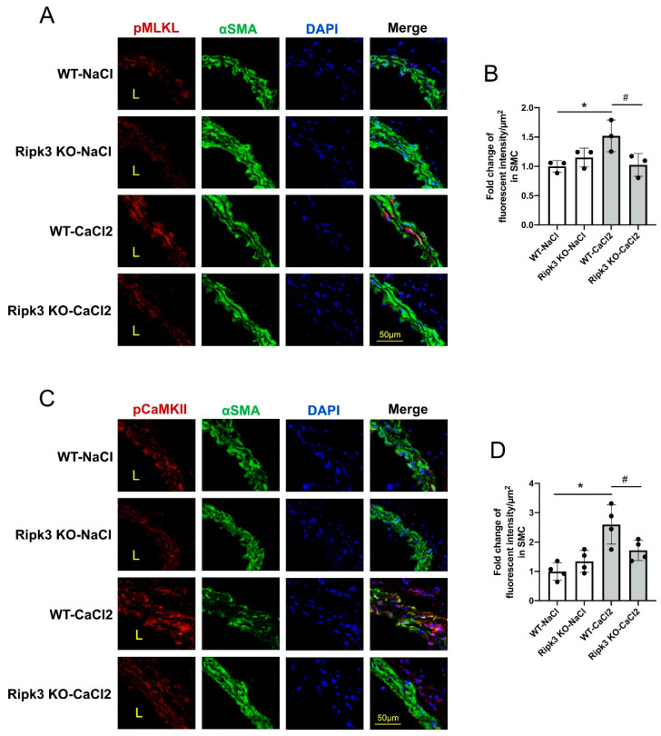
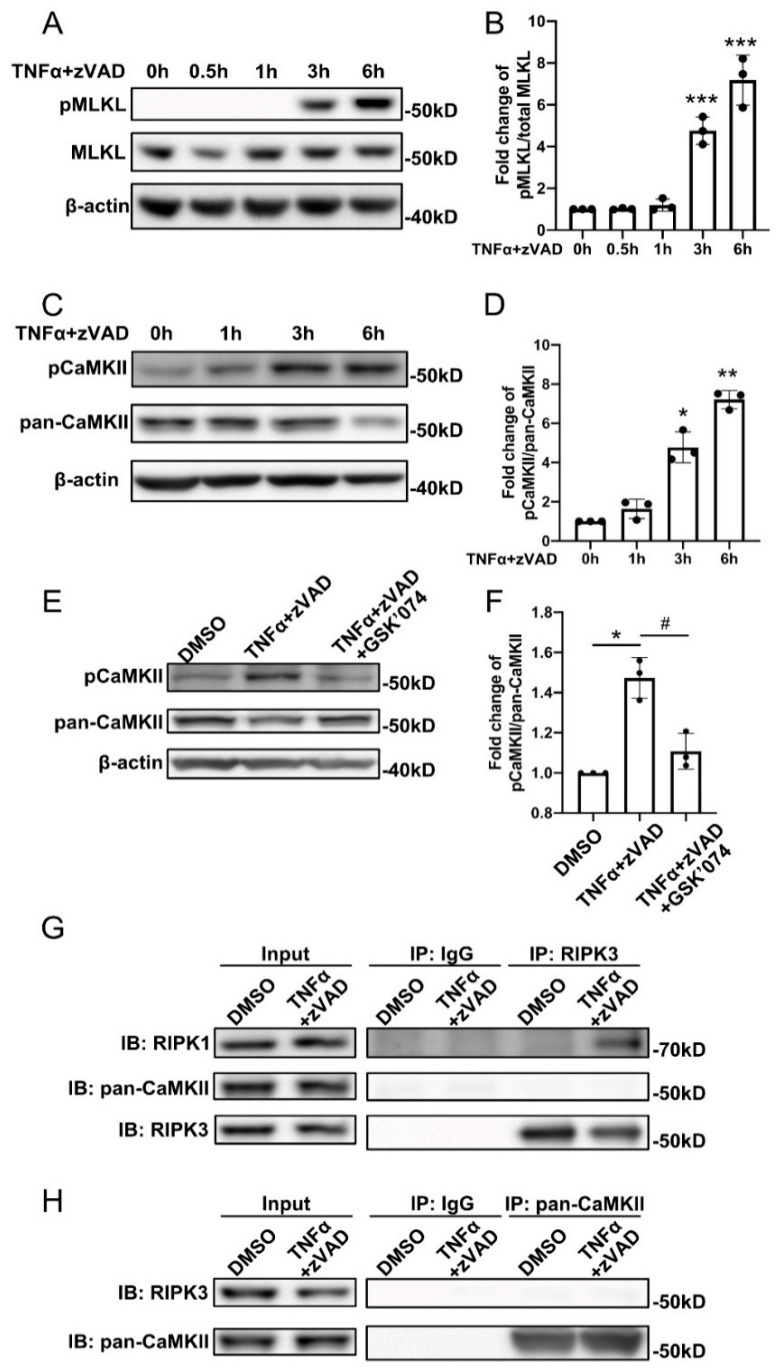
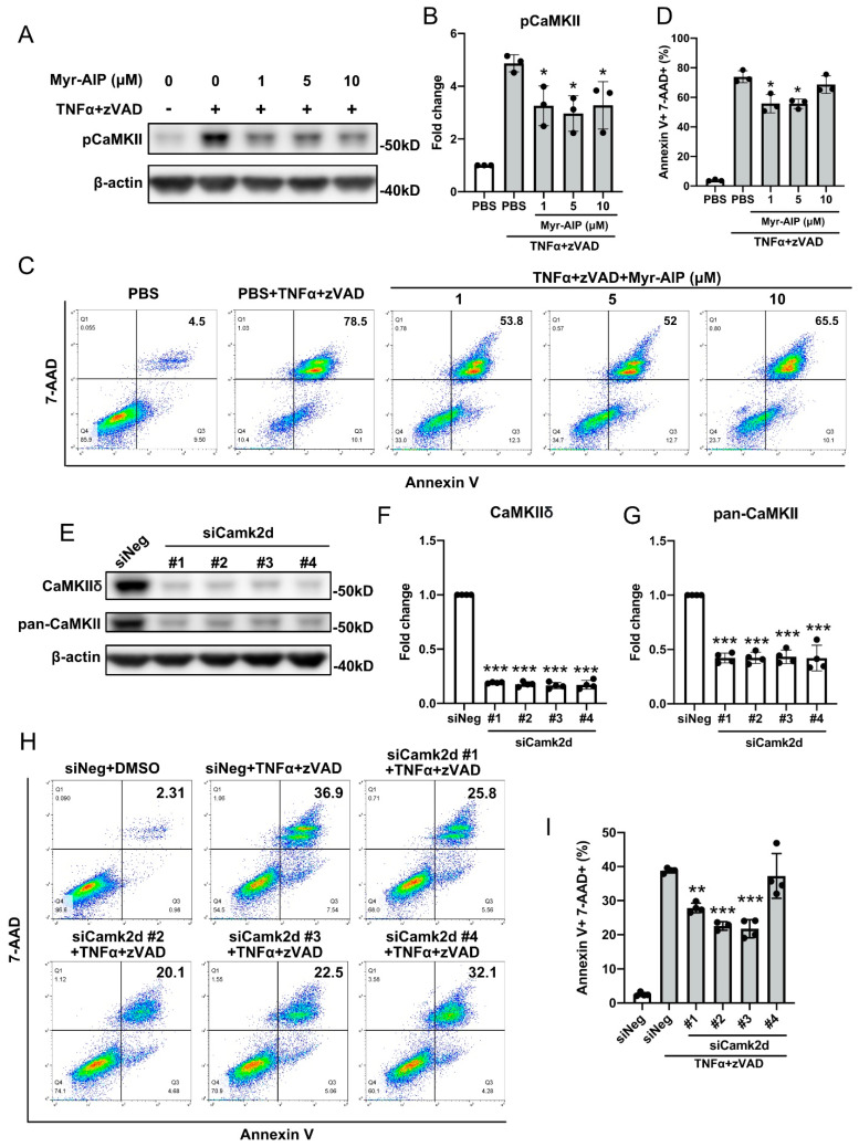
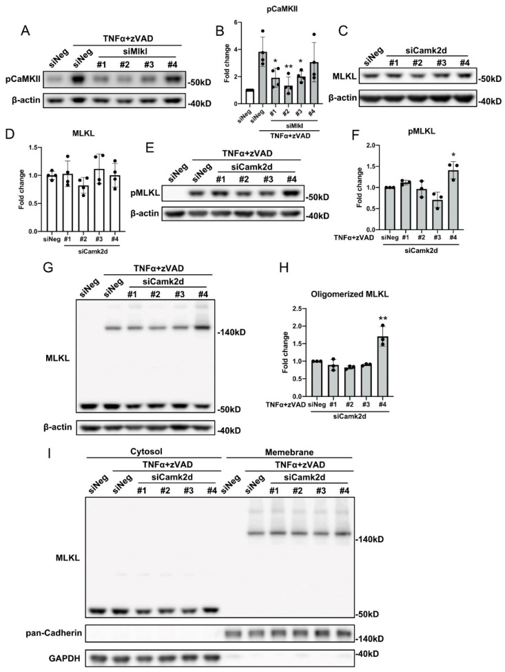
Receptor interacting protein kinase 3 (RIPK3)-mediated smooth muscle cell (SMC) necroptosis has been shown to contribute to the pathogenesis of abdominal aortic aneurysms (AAAs). However, the signaling steps downstream from RIPK3 during SMC necroptosis remain unknown. In this study, the roles of mixed lineage kinase domain-like pseudokinase (MLKL) and calcium/calmodulin-dependent protein kinase II (CaMKII) in SMC necroptosis were investigated. We found that both MLKL and CaMKII were phosphorylated in SMCs in a murine CaCl2-driven model of AAA and that Ripk3 deficiency reduced the phosphorylation of MLKL and CaMKII. In vitro, mouse aortic SMCs were treated with tumor necrosis factor α (TNFα) plus Z-VAD-FMK (zVAD) to induce necroptosis. Our data showed that both MLKL and CaMKII were phosphorylated after TNFα plus zVAD treatment in a time-dependent manner. SiRNA silencing of Mlkl-diminished cell death and administration of the CaMKII inhibitor myristoylated autocamtide-2-related inhibitory peptide (Myr-AIP) or siRNAs against Camk2d partially inhibited necroptosis. Moreover, knocking down Mlkl decreased CaMKII phosphorylation, but silencing Camk2d did not affect phosphorylation, oligomerization, or trafficking of MLKL. Together, our results indicate that both MLKL and CaMKII are involved in RIPK3-mediated SMC necroptosis, and that MLKL is likely upstream of CaMKII in this process.

Keywords: CaMKII; MLKL; abdominal aortic aneurysm; necroptosis; smooth muscle cells.


Conflict of interest statement

The authors declare no conflict of interest.

Figures

Figure 1
Figure 1
Elevated phosphorylation of MLKL and CaMKII in a murine CaCl2-induced AAA model. Ripk3 wildtype (WT) and knockout (KO) mice were subjected to AAA induction by perivascular treatment with CaCl2 or NaCl (sham group). Four days after AAA induction, treated abdominal aortic segments were harvested for cross sections. (A,C) Anti-alpha smooth muscle actin (green) was used to identify medial smooth muscle cells and DAPI was used to stain nuclei. Representative images of immunostaining of phospho-MLKL Ser345 (red) are shown in panel A and phospho-CaMKII Thr287 (red) in panel C. (B,D) Quantification of fluorescent intensity of phospho-MLKL Ser345 (B) and phospho-CaMKII Thr287 (D) is presented relative to the medial layer area. n = 3 and 4 for each group in (B) and (D), respectively. Data were presented as mean ± SD. One-way ANOVA was performed in (B) and (D). * p < 0.05, compared with WT NaCl group; # p < 0.05, compared with WT CaCl2 group.
Figure 2
Figure 2
Both MLKL and CaMKII are activated during SMC necroptosis. (A,C) Mouse aortic smooth muscle cells (MOVAS) were treated with 30 ng/mL TNFα plus 60 µM zVAD for the indicated time period. Cells were lysed in RIPA buffer and analyzed by Western blotting with the indicated antibodies. (B) Quantification of (A). n = 3 for each timepoint. (D) Quantification of (C). n = 3 for each timepoint. (E) MOVAS were treated with 30 ng/mL TNFα plus 60 µM zVAD for 6 h in the presence or absence of 10 nM GSK’074. Cells were lysed in RIPA buffer and analyzed by Western blotting with the indicated antibodies. (F) Quantification of (E). n = 3. (G,H) MOVAS cells were treated with 30 ng/mL TNFα plus 60 μM zVAD for 6 h. Cell lysates were immunoprecipitated with anti-IgG, anti-RIPK3 (G), or anti-pan CaMKII (H) antibodies followed by Western blotting analysis with the indicated antibodies. Data were presented as mean ± SD of at least three independent experiments. One-way ANOVA was performed in (B,D,F). * p < 0.05, ** p < 0.01, *** p < 0.001. In (F), * p < 0.05, compared with DMSO group; # p < 0.05, compared with TNFα+zVAD group.
Figure 3
Figure 3
MLKL plays an essential role in SMC necroptosis. MOVAS cells were transfected with siRNAs against Mlkl for 48 h. (A,B) Validation of siRNA knockdown by Western blotting. (C) Following Mlkl knockdown, MOVAS cells were treated with 30 ng/mL TNFα plus 60 µM zVAD for 6 h. Cells were then stained with 7-AAD and Annexin V, and analyzed by flow cytometry. Necrotic cells were identified as 7-AAD+ Annexin V+. (D) Quantification of (C). Data were presented as mean ± SD of at least three independent experiments. One-way ANOVA was performed in (B,D). *** p < 0.001.
Figure 4
Figure 4
CaMKII is partially required for SMC necroptosis. (AD) MOVAS cells were pretreated with indicated doses of CaMKII inhibitor myristoylated autocamtide-2-related inhibitory peptide (Myr-AIP) for 1 h, followed by 6 h of treatment with 30 ng/mL TNFα plus 60 µM zVAD. (A) Cells were lysed in RIPA buffer and subjected to Western blotting analysis with the indicated antibodies. (C) Cells were stained with 7-AAD and Annexin V, and subjected to flow cytometry analysis. Necrotic cells were identified as 7-AAD+ Annexin V+. (B) was quantification of (A). (D) was quantification of (C). (E) MOVAS cells were transfected with siRNAs against Camk2d for 48 h. Cells were lysed in RIPA buffer and subjected to Western blotting analysis with the indicated antibodies. (F,G) Quantification of (E). (H) MOVAS cells were transfected with siRNAs against Camk2d for 48 h, then treated with 30 ng/mL TNFα plus 60 µM zVAD for 6 h. Cells were then stained with 7-AAD and Annexin V, and subjected to flow cytometry analysis. Necrotic cells were identified as 7-AAD+ Annexin V+. (I) Quantification of (H). Data were presented as mean ± SD of at least three independent experiments. One-way ANOVA was performed in (B,D,F,G,I). * p < 0.05, ** p < 0.01, *** p < 0.001 compared with TNFα plus zVAD treated group (B,D,I) or scrambled siRNA-treated group (F,G).
Figure 5
Figure 5
CaMKII activation requires MLKL. (A) MOVAS cells were transfected with different siRNAs against Mlkl for 48 h, then treated with 30 ng/mL TNFα plus 60 µM zVAD for 6 h. Cells were lysed in RIPA buffer and subjected to Western blotting analysis with the indicated antibodies. (B) Quantification of (A). (C) MOVAS cells were transfected with siRNAs against Camk2d for 48 h, cells were lysed in RIPA buffer and subjected to Western blotting analysis with the indicated antibodies. (D) Quantification of (C). (EI) MOVAS cells were transfected with siRNAs against Camk2d for 48 h, then treated with 30 ng/mL TNFα plus 60 µM zVAD for 6 h. (E,G) Cells were lysed in RIPA buffer (E) or 1% digitonin (G) and were subjected to Western blotting analysis with the indicated antibodies. (I) Cytosol and membrane fractions were subjected to Western blotting analysis with the indicated antibodies. (F) Quantification of (E). (H) Quantification of (G). Data were presented as mean ± SD of at least three independent experiments. One-way ANOVA was performed in (B,D,F,H). * p < 0.05, ** p < 0.01 compared with TNFα plus zVAD-treated group (B,F,H).

References

    1. Quintana R.A., Taylor W.R. Cellular Mechanisms of Aortic Aneurysm Formation. Circ. Res. 2019;124:607–618. doi: 10.1161/CIRCRESAHA.118.313187. - DOI - PMC - PubMed
    1. Wang Q., Liu Z., Ren J., Morgan S., Assa C., Liu B. Receptor-interacting protein kinase 3 contributes to abdominal aortic aneurysms via smooth muscle cell necrosis and inflammation. Circ. Res. 2015;116:600–611. doi: 10.1161/CIRCRESAHA.116.304899. - DOI - PMC - PubMed
    1. Shan B., Pan H., Najafov A., Yuan J. Necroptosis in development and diseases. Genes Dev. 2018;32:327–340. doi: 10.1101/gad.312561.118. - DOI - PMC - PubMed
    1. Wang Q., Zhou T., Liu Z., Ren J., Phan N., Gupta K., Stewart D.M., Morgan S., Assa C., Kent K.C., et al. Inhibition of Receptor-Interacting Protein Kinase 1 with Necrostatin-1s ameliorates disease progression in elastase-induced mouse abdominal aortic aneurysm model. Sci. Rep. 2017;7:42159. doi: 10.1038/srep42159. - DOI - PMC - PubMed
    1. Zhou T., Wang Q., Phan N., Ren J., Yang H., Feldman C.C., Feltenberger J.B., Ye Z., Wildman S.A., Tang W., et al. Identification of a novel class of RIP1/RIP3 dual inhibitors that impede cell death and inflammation in mouse abdominal aortic aneurysm models. Cell Death Dis. 2019;10:226. doi: 10.1038/s41419-019-1468-6. - DOI - PMC - PubMed

Publication types

MeSH terms

Substances

LinkOut - more resources