

Betulinic Acid Protects from Ischemia-Reperfusion Injury in the Mouse Retina
- PMID: 34572088
- PMCID: PMC8469383
- DOI: 10.3390/cells10092440
Betulinic Acid Protects from Ischemia-Reperfusion Injury in the Mouse Retina
Abstract
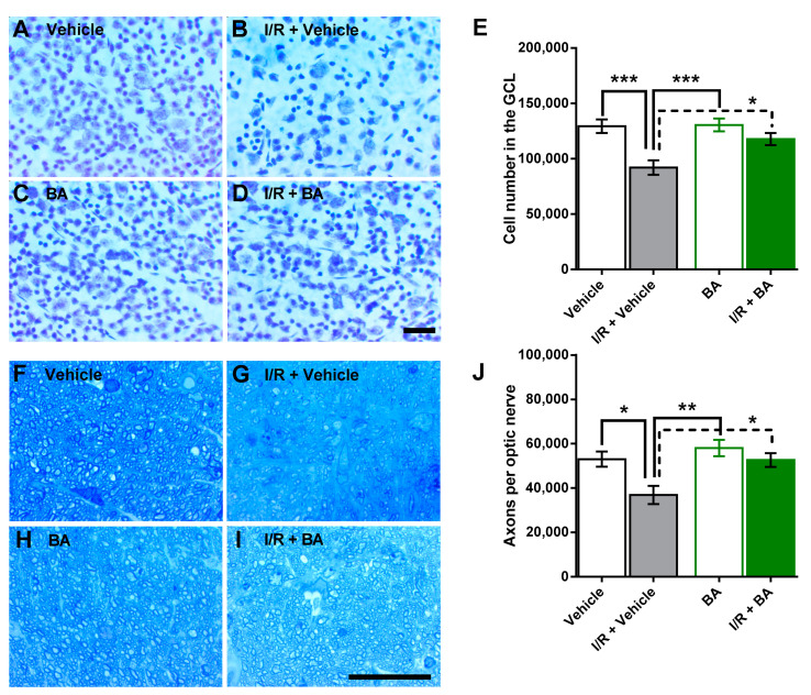
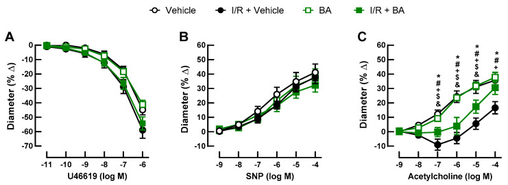
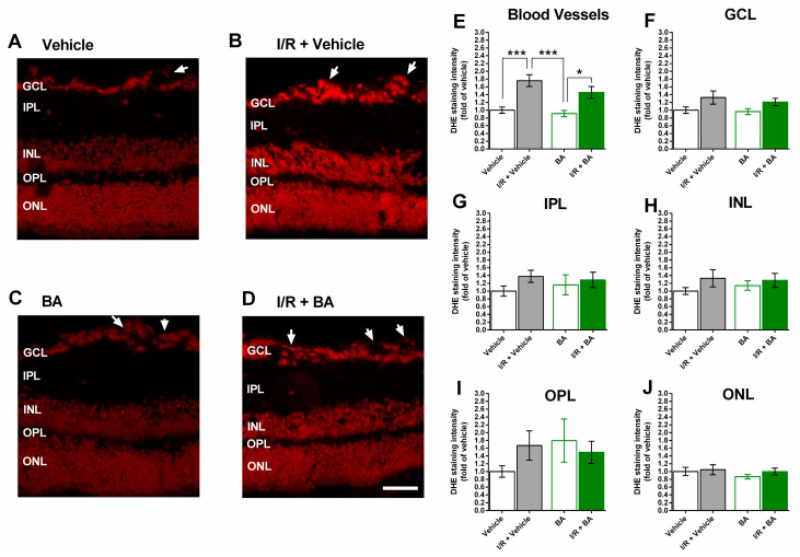
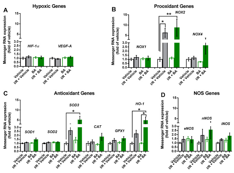
Ischemia/reperfusion (I/R) events are involved in the pathophysiology of numerous ocular diseases. The purpose of this study was to test the hypothesis that betulinic acid protects from I/R injury in the mouse retina. Ocular ischemia was induced in mice by increasing intraocular pressure (IOP) to 110 mm Hg for 45 min, while the fellow eye served as a control. One group of mice received betulinic acid (50 mg/kg/day p.o. once daily) and the other group received the vehicle solution only. Eight days after the I/R event, the animals were killed and the retinal wholemounts and optic nerve cross-sections were prepared and stained with cresyl blue or toluidine blue, respectively, to count cells in the ganglion cell layer (GCL) of the retina and axons in the optic nerve. Retinal arteriole responses were measured in isolated retinas by video microscopy. The levels of reactive oxygen species (ROS) were assessed in retinal cryosections and redox gene expression was determined in isolated retinas by quantitative PCR. I/R markedly reduced cell number in the GCL and axon number in the optic nerve of the vehicle-treated mice. In contrast, only a negligible reduction in cell and axon number was observed following I/R in the betulinic acid-treated mice. Endothelial function was markedly reduced and ROS levels were increased in retinal arterioles of vehicle-exposed eyes following I/R, whereas betulinic acid partially prevented vascular endothelial dysfunction and ROS formation. Moreover, betulinic acid boosted mRNA expression for the antioxidant enzymes SOD3 and HO-1 following I/R. Our data provide evidence that betulinic acid protects from I/R injury in the mouse retina. Improvement of vascular endothelial function and the reduction in ROS levels appear to contribute to the neuroprotective effect.
Keywords: arterioles; betulinic acid; ischemia-reperfusion injury; reactive oxygen species; retina.
Conflict of interest statement
The authors declare no conflict of interest. The funders had no role in the design of the study; in the collection, analyses, or interpretation of data; in the writing of the manuscript, or in the decision to publish the results.
Figures
References
-
- Feltgen N., Neubauer A., Jurklies B., Schmoor C., Schmidt D., Wanke J., Maier-Lenz H., Schumacher M. Multicenter study of the European Assessment Group for Lysis in the Eye (EAGLE) for the treatment of central retinal artery occlusion: Design issues and implications. EAGLE Study report no. 1: EAGLE Study report no. 1. Graefes Arch. Clin. Exp. Ophthalmol. 2006;244:950–956. doi: 10.1007/s00417-005-0140-2. - DOI - PubMed
-
- Schumacher M., Schmidt D., Jurklies B., Gall C., Wanke I., Schmoor C., Maier-Lenz H., Solymosi L., Brueckmann H., Neubauer A.S., et al. Central Retinal Artery Occlusion: Local Intra-arterial Fibrinolysis versus Conservative Treatment, a Multicenter Randomized Trial. Ophthalmology. 2010;117:1367–1375.e1. doi: 10.1016/j.ophtha.2010.03.061. - DOI - PubMed
Publication types
MeSH terms
Substances
LinkOut - more resources
Full Text Sources
Miscellaneous
