

The Role of Mitochondria in Oocyte Maturation
- PMID: 34572133
- PMCID: PMC8469615
- DOI: 10.3390/cells10092484
The Role of Mitochondria in Oocyte Maturation
Abstract
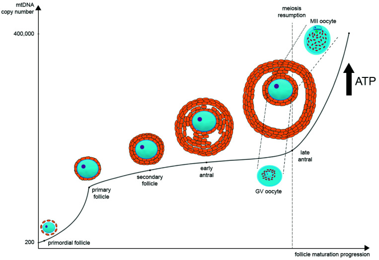
With the nucleus as an exception, mitochondria are the only animal cell organelles containing their own genetic information, called mitochondrial DNA (mtDNA). During oocyte maturation, the mtDNA copy number dramatically increases and the distribution of mitochondria changes significantly. As oocyte maturation requires a large amount of ATP for continuous transcription and translation, the availability of the right number of functional mitochondria is crucial. There is a correlation between the quality of oocytes and both the amount of mtDNA and the amount of ATP. Suboptimal conditions of in vitro maturation (IVM) might lead to changes in the mitochondrial morphology as well as alternations in the expression of genes encoding proteins associated with mitochondrial function. Dysfunctional mitochondria have a lower ability to counteract reactive oxygen species (ROS) production which leads to oxidative stress. The mitochondrial function might be improved with the application of antioxidants and significant expectations are laid on the development of new IVM systems supplemented with mitochondria-targeted reagents. Different types of antioxidants have been tested already on animal models and human rescue IVM oocytes, showing promising results. This review focuses on the recent observations on oocytes' intracellular mitochondrial distribution and on mitochondrial genomes during their maturation, both in vivo and in vitro. Recent mitochondrial supplementation studies, aiming to improve oocyte developmental potential, are summarized.
Keywords: IVM; mitochondria distribution; mitochondrial supplementation reagents; mtDNA copy number; oocyte maturation.
Conflict of interest statement
The authors declare no conflict of interest.
Figures
References
-
- Practice Committees of the American Society for Reproductive Medicine, the Society of Reproductive Biologists and Technologists, and the Society for Assisted Reproductive Technology In Vitro Maturation: A Committee Opinion. Fertil. Steril. 2021;115:298–304. doi: 10.1016/j.fertnstert.2020.11.018. - DOI - PubMed
-
- Walls M.L., Hunter T., Ryan J.P., Keelan J.A., Nathan E., Hart R.J. In Vitro Maturation as an Alternative to Standard in Vitro Fertilization for Patients Diagnosed with Polycystic Ovaries: A Comparative Analysis of Fresh, Frozen and Cumulative Cycle Outcomes. Hum. Reprod. 2015;30:88–96. doi: 10.1093/humrep/deu248. - DOI - PubMed
Publication types
MeSH terms
Substances
Grants and funding
LinkOut - more resources
Full Text Sources
Medical
Research Materials
