. 2021 Aug 26;9(9):1089.
doi: 10.3390/biomedicines9091089.

Wharton's Jelly-Derived Mesenchymal Stem Cells Reduce Fibrosis in a Mouse Model of Duchenne Muscular Dystrophy by Upregulating microRNA 499

Affiliations

Wharton's Jelly-Derived Mesenchymal Stem Cells Reduce Fibrosis in a Mouse Model of Duchenne Muscular Dystrophy by Upregulating microRNA 499

Sang Eon Park et al. Biomedicines. .

Abstract

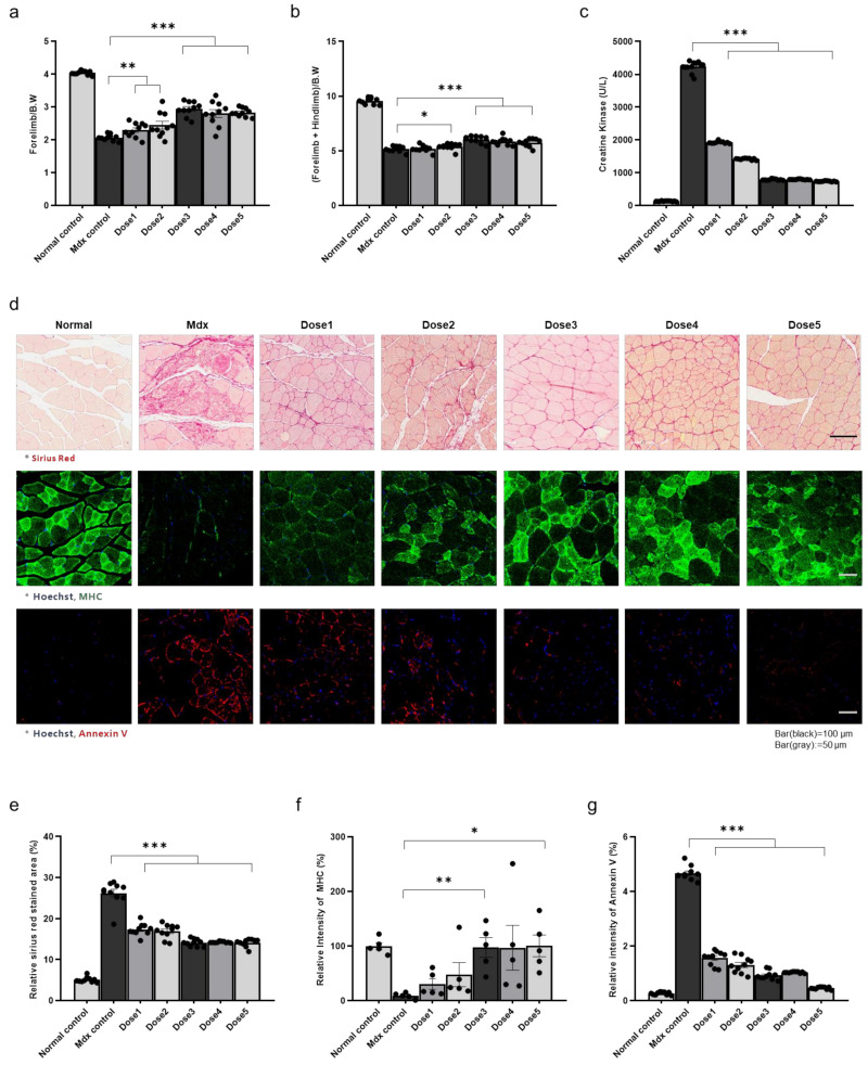
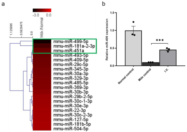
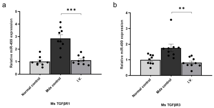
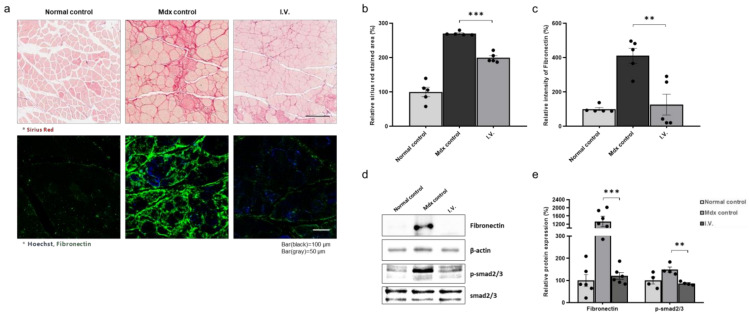
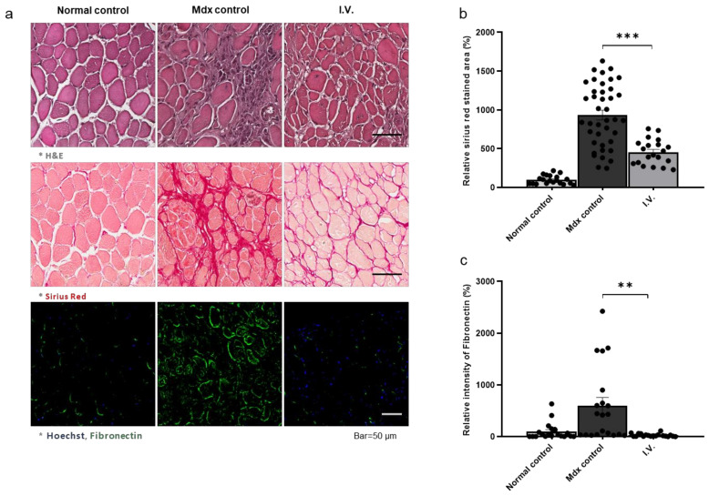
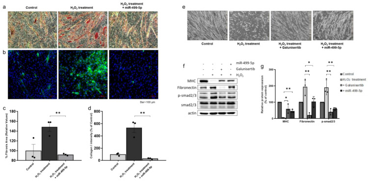
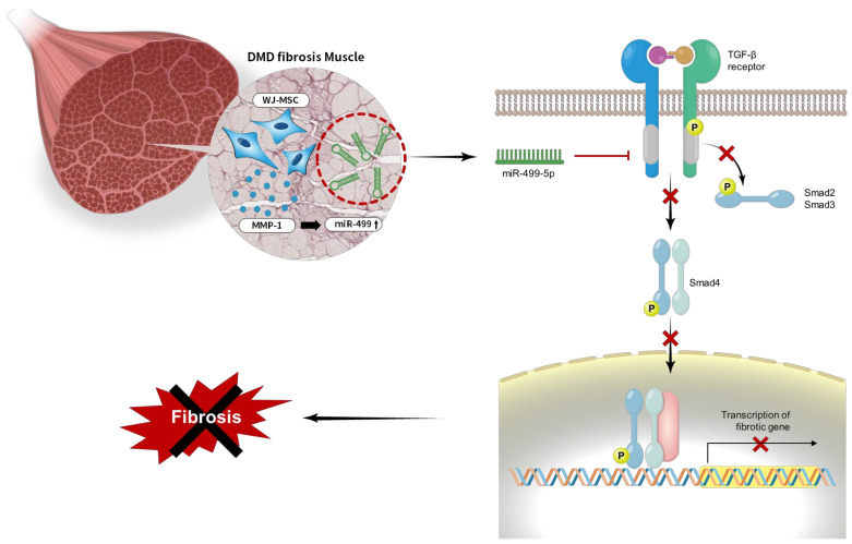
The aim of this study was to evaluate the therapeutic effects and mechanisms of Wharton's jelly-derived mesenchymal stem cells (WJ-MSCs) in an animal model of Duchenne muscular dystrophy (DMD). Mdx mice (3-5 months old) were administered five different doses of WJ-MSCs through their tail veins. A week after injection, grip strength measurements, creatine kinase (CK) assays, immunohistochemistry, and western blots were performed for comparison between healthy mice, mdx control mice, and WJ-MSC-injected mdx mice. WJ-MSCs exerted dose-dependent multisystem therapeutic effects in mdx mice, by decreasing CK, recovering normal behavior, regenerating muscle, and reducing apoptosis and fibrosis in skeletal muscle. We also confirmed that miR-499-5p is significantly downregulated in mdx mice, and that intravenous injection of WJ-MSCs enhanced its expression, leading to anti-fibrotic effects via targeting TGFβR 1 and 3. Thus, WJ-MSCs may represent novel allogeneic "off-the-shelf" cellular products for the treatment of DMD and possibly other muscle disorders.

Keywords: Duchenne muscular dystrophy; Wharton’s jelly-derived mesenchymal stem cell; microRNA-499-5p; skeletal muscle fibrosis.


Conflict of interest statement

The authors declare no conflict of interest. The funders had no role in the design of the study; in the collection, analyses, or interpretation of data; in the writing of the manuscript, or in the decision to publish the results.

Figures

Figure 1
Figure 1
Therapeutic effects of Wharton’s jelly-derived mesenchymal stem cells (WJ-MSCs) in the skeletal muscle of mdx mice (a) Forelimb grip strength, (b) forelimb + hindlimb grip strength, (c) creatine kinase (CK) levels, (d) fibrotic areas, and myosin heavy chain and annexin V expression levels were measured in control, untreated mdx mice, and WJ-MSC-treated mdx mice. (eg) Quantitative measurements were performed in ImageJ. The data are expressed as the mean ± SEM. Bars with different superscripts indicate significantly different values (* p < 0.05, ** p < 0.01, *** p < 0.001).
Figure 2
Figure 2
miR-499-5p is downregulated in mdx mice compared to healthy mice (a) Heatmap depicting the levels of miRNAs commonly expressed in humans and mice in mdx mice and healthy mice. (b) Relative miR-499-5p expression levels in the skeletal muscles of each group. miR-26a-5p was used as an endogenous control. The data are expressed as the mean ± SEM. Bars with different superscripts indicate significantly different (*** p < 0.001).
Figure 3
Figure 3
Transforming growth factor β receptor (TGFβR)1 and TGFβR3 are direct targets of miR-499-5p (a) Relative TGFβR1 expression levels were measured in skeletal muscles from each group. Glyceraldehyde 3-phosphate dehydrogenase (GAPDH) was used as an endogenous control, and data are expressed as the mean ± the SEM. Bars with different superscripts indicate significantly different values. (b) Relative TGFβR3 expression levels were measured in the skeletal muscle of each group. GAPDH was used as an endogenous control, and data are expressed as mean ± SEM (** p < 0.01, *** p < 0.001).
Figure 4
Figure 4
Anti-fibrotic effect of Wharton’s jelly-derived mesenchymal stem cells in the skeletal muscle of mdx mice (a) Representative images of Sirius Red staining for fibrosis evaluation and immunohistochemistry for fibronectin detection. (b,c) Quantitative measurements were performed in ImageJ, and the data are expressed as the mean ± SEM. Bars with different superscripts indicate significantly different values. (d) Representative western blot for fibronectin and p-smad2/3. β-actin was used as a loading control. Phospho-smad2/3 was normalized to total smad2/3. (e) Quantification of fibronectin and phospho-smad2/3 protein levels. The data are expressed as the mean ± SEM. Bars with different superscripts indicate significantly different values (** p < 0.01, *** p < 0.001).
Figure 5
Figure 5
Antifibrotic effect of Wharton’s jelly-derived mesenchymal stem cells in the diaphragm of mdx mice (a) Representative images of H&E and Sirius red staining and immunohistochemistry for fibronectin detection. (b,c) Quantitative measurements were performed in ImageJ, and the data are expressed as the mean ± SEM. Bars with different superscripts indicate significantly different values (** p < 0.01, *** p < 0.001).
Figure 6
Figure 6
Effects of miR-499-5p on an H2O2-induced myotube fibrosis model (a,c) Fibrosis was assessed as the percentage of the area positive for Sirius Red staining. Quantitative measurements were performed in ImageJ, and data are expressed as the mean ± SEM. Bars with different superscripts indicate significantly different values (** p < 0.01). Scale bars: 100 μm. (b,d) Immunocytochemistry to detect collagen I deposition in myotubes. Quantitative measurements were performed in ImageJ, and data are expressed as mean ± SEM. Bars with different superscripts indicate significantly different values (** p < 0.01). Scale bars: 100 μm. (e) Differentiated myotubes were treated with 2 mM H2O2 in the absence or presence of galunisertib and miR-499-5p mimic. Administration of galunisertib and miR-499-5p mimic induced recovery of myotubes damaged by H2O2 treatment (bar = 100 µm). (f) Representative western blots for myosin heavy chain, fibronectin, and phospho-smad2/3. β-actin was used as a loading control. Phospho-smad2/3 was normalized to total smad2/3. (g) Quantification of myosin heavy chain, fibronectin and phospho-smad2/3 protein levels. Data are expressed as the mean ± SEM. Bars with different superscripts indicate significantly different values (* p < 0.05, ** p < 0.01).
Figure 7
Figure 7
A model of miR-499-dependent anti-fibrotic signaling pathway. Overall proposed model of the anti-fibrotic mechanism of miR-499-5p in skeletal muscles in Duchenne muscular dystrophy (DMD). The activation of TGF β signaling pathway induces fibrosis. Our findings suggest that MMP-1 secreted from WJ-MSCs increases miR-499 expression level. Increased miR-499-5p down-regulates to the TGFβ receptor I and blocks smad2/3 phosphorylation, leading to fibrotic muscle recovery by the inactivated TGFβ signaling pathway (formula image: block, formula image: inactivation).

References

    1. Emery A.E.H. The muscular dystrophies. Lancet. 2002;359:687–695. doi: 10.1016/S0140-6736(02)07815-7. - DOI - PubMed
    1. Nowak K.J., Davies K.E. Duchenne muscular dystrophy and dystrophin: Pathogenesis and opportunities for treatment. EMBO Rep. 2004;5:872–876. doi: 10.1038/sj.embor.7400221. - DOI - PMC - PubMed
    1. De Paepe B. Progressive Skeletal Muscle Atrophy in Muscular Dystrophies: A Role for Toll-like Receptor-Signaling in Disease Pathogenesis. Int. J. Mol. Sci. 2020;21:4440. doi: 10.3390/ijms21124440. - DOI - PMC - PubMed
    1. Grady R.M., Teng H., Nichol M.C., Cunningham J.C., Wilkinson R.S., Sanes J.R. Skeletal and Cardiac Myopathies in Mice Lacking Utrophin and Dystrophin: A Model for Duchenne Muscular Dystrophy. Cell. 1997;90:729–738. doi: 10.1016/S0092-8674(00)80533-4. - DOI - PubMed
    1. Eagle M., Baudouin S.V., Chandler C., Giddings D.R., Bullock R., Bushby K. Survival in Duchenne muscular dystrophy: Improvements in life expectancy since 1967 and the impact of home nocturnal ventilation. Neuromuscul. Disord. 2002;12:926–929. doi: 10.1016/S0960-8966(02)00140-2. - DOI - PubMed