

High PD-L1 Expression on Tumor Cells Indicates Worse Overall Survival in Advanced Oral Squamous Cell Carcinomas of the Tongue and the Floor of the Mouth but Not in Other Oral Compartments
- PMID: 34572318
- PMCID: PMC8471659
- DOI: 10.3390/biomedicines9091132
High PD-L1 Expression on Tumor Cells Indicates Worse Overall Survival in Advanced Oral Squamous Cell Carcinomas of the Tongue and the Floor of the Mouth but Not in Other Oral Compartments
Abstract
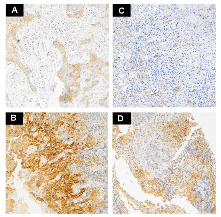
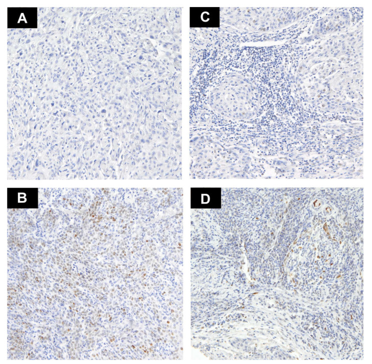
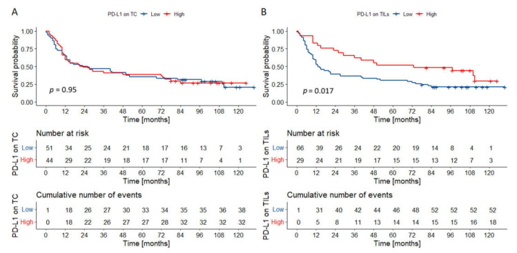
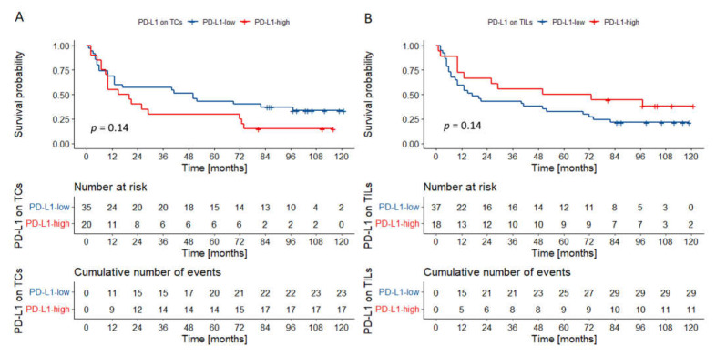
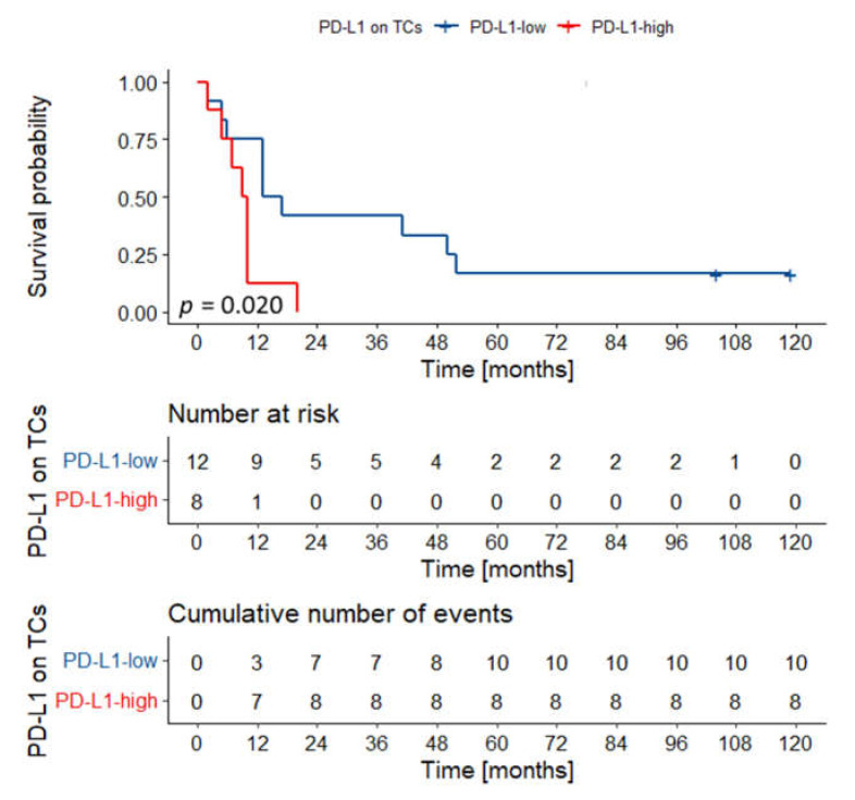
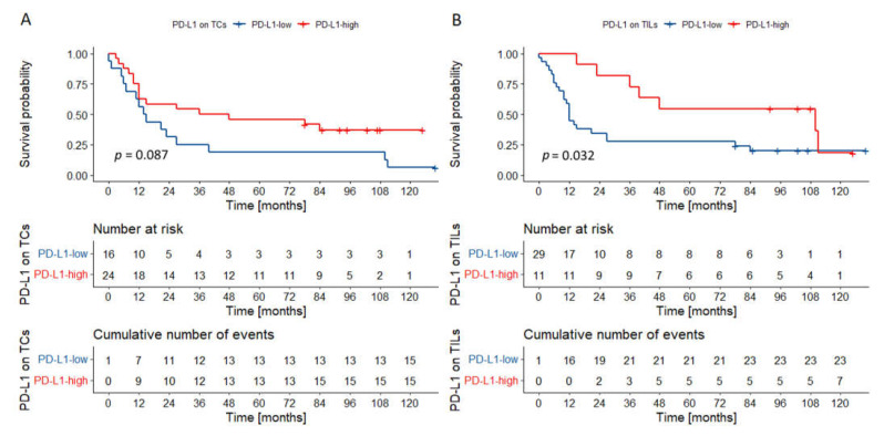
The markers of the tumor microenvironment (TME) are promising prognostic and predictive factors in oral squamous cell carcinoma (OSCC). The current study aims to analyze the immunohistochemical expression of programmed cell death-ligand 1 (PD-L1) and interleukin-33 (IL-33) in a cohort of 95 chemonaïve OSCCs. PD-L1 and IL-33 were assessed separately in tumor cells (TCs) and tumor-infiltrating lymphocytes (TILs). High PD-L1 expression in TILs was associated with better overall survival (OS) in univariate analysis. Tumors localized in the floor of the oral cavity and tongue tended to have a lower percentage of PD-L1-positive TCs when compared to other locations. PD-L1 expression on TCs had no prognostic significance when the whole cohort was analyzed. However, along with the T descriptor (TNM 8th), it was included in the multivariable model predicting death in carcinomas of the floor of the oral cavity and tongue (HR = 2.51, 95% CI = 1.97-5.28). In other locations, only nodal status was identified as an independent prognostic factor in multivariate analysis (HR = 0.24, 95% CI = 0.08-0.70). Expression of IL-33 had no impact on survival, but it was differently expressed in various locations. In conclusion, the prognostic significance of PD-L1 in oral cancer depends on the tumor site and type of cell expressing immune checkpoint receptor (TCs vs. TILs).
Keywords: IL-33; PD-L1; immunohistochemistry; interleukin 33; oral squamous cell carcinoma; prognosis; programmed cell death-ligand 1.
Conflict of interest statement
The authors declare no conflict of interest.
Figures
Similar articles
-
Negative prognostic behaviour of PD-L1 expression in tongue and larynx squamous cell carcinoma and its significant predictive power in combination with PD-1 expression on TILs.BMC Immunol. 2024 Jan 16;25(1):7. doi: 10.1186/s12865-024-00597-0. BMC Immunol. 2024. PMID: 38229027 Free PMC article.
-
IL-12RB1: a novel immune prognostic biomarker for oral squamous cell carcinoma and linked to PD-1/PD-L1 expression in the tumor immune microenvironment.Ann Transl Med. 2022 Feb;10(3):144. doi: 10.21037/atm-21-6915. Ann Transl Med. 2022. PMID: 35284546 Free PMC article.
-
Clinical significance of programmed cell death-ligand expression in small bowel adenocarcinoma is determined by the tumor microenvironment.World J Gastroenterol. 2023 Oct 28;29(40):5566-5581. doi: 10.3748/wjg.v29.i40.5566. World J Gastroenterol. 2023. PMID: 37970475 Free PMC article.
-
Relationship of programmed death ligand-1 expression with clinicopathological features and prognosis in patients with oral squamous cell carcinoma: A meta-analysis.Arch Oral Biol. 2020 Jun;114:104717. doi: 10.1016/j.archoralbio.2020.104717. Epub 2020 Apr 11. Arch Oral Biol. 2020. PMID: 32344357 Review.
-
Prognostic role of different PD-L1 expression patterns and tumor-infiltrating lymphocytes in high-grade serous ovarian cancer: a systematic review and meta-analysis.Front Immunol. 2023 Aug 15;14:1234894. doi: 10.3389/fimmu.2023.1234894. eCollection 2023. Front Immunol. 2023. PMID: 37654479 Free PMC article.
Cited by
-
VISTA H-Score Is Significantly Associated with a 5-Year DFS Rate in Oral Squamous Cell Carcinoma.J Clin Med. 2023 Feb 17;12(4):1619. doi: 10.3390/jcm12041619. J Clin Med. 2023. PMID: 36836154 Free PMC article.
-
High PD-L1 expression on immune cells along with increased density of tumor-infiltrating lymphocytes predicts a favorable survival outcome for patients with loco-regionally advanced head and neck cancer: early results from a prospective study.Front Oncol. 2024 Apr 4;14:1346793. doi: 10.3389/fonc.2024.1346793. eCollection 2024. Front Oncol. 2024. PMID: 38638854 Free PMC article.
-
Programmed Cell Death Ligand 1 Expression on Immune Cells and Survival in Patients With Nonmetastatic Head and Neck Cancer: A Systematic Review and Meta-analysis.JAMA Netw Open. 2023 Mar 1;6(3):e236324. doi: 10.1001/jamanetworkopen.2023.6324. JAMA Netw Open. 2023. PMID: 37000447 Free PMC article.
-
PD-L1 in oral squamous cell carcinoma: A key biomarker from the laboratory to the bedside.Clin Exp Dent Res. 2022 Jun;8(3):690-698. doi: 10.1002/cre2.590. Epub 2022 May 20. Clin Exp Dent Res. 2022. PMID: 35593124 Free PMC article. Review.
-
Pathological examination of factors involved in PD-L1 expression in patients with oral tongue squamous cell carcinoma.Maxillofac Plast Reconstr Surg. 2024 Aug 8;46(1):31. doi: 10.1186/s40902-024-00441-w. Maxillofac Plast Reconstr Surg. 2024. PMID: 39115623 Free PMC article.
References
-
- Feller L., Lemmer J. Oral Squamous Cell Carcinoma: Epidemiology, Clinical Presentation and Treatment. J. Cancer Ther. 2012;03:263–268. doi: 10.4236/jct.2012.34037. - DOI
-
- Starzyńska A., Sejda A., Adamski Ł., Adamska P., Pęksa R., Sakowicz-Burkiewicz M., Wychowański P., Jereczek-Fossa B.A. The B7 family molecules in oral squamous cell carcinoma: A systematic review. Part I: B7-H1 (PD-L1) and B7-DC (PD-L2) Adv. Dermatol. Allergol. 2020;37 doi: 10.5114/ada.2020.98522. - DOI - PMC - PubMed
-
- Starzyńska A., Sejda A., Adamski Ł., Adamska P., Pęksa R., Sakowicz-Burkiewicz M., Wychowański P., Jereczek-Fossa B.A. The B7 family molecules in oral squamous cell carcinoma: A systematic review. Part II: B7-1, B7-2, B7-H2, B7-H3, B7-H4, B7-H5 (VISTA), B7-H6 and B7-H7. Adv. Dermatol. Allergol. 2020;37 doi: 10.5114/ada.2020.98523. - DOI - PMC - PubMed
-
- Binnewies M., Roberts E.W., Kersten K., Chan V., Fearon D.F., Merad M., Coussens L.M., Gabrilovich D.I., Ostrand-Rosenberg S., Hedrick C.C., et al. Understanding the tumor immune microenvironment (TIME) for effective therapy. Nat. Med. 2018;24:541–550. doi: 10.1038/s41591-018-0014-x. - DOI - PMC - PubMed
LinkOut - more resources
Full Text Sources
Research Materials
