Review
. 2021 Sep 10;9(9):1188.
doi: 10.3390/biomedicines9091188.

The Role of Smoothened-Dependent and -Independent Hedgehog Signaling Pathway in Tumorigenesis

Affiliations
Review

The Role of Smoothened-Dependent and -Independent Hedgehog Signaling Pathway in Tumorigenesis

Jian Yi Chai et al. Biomedicines. .

Abstract

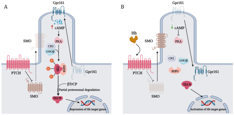
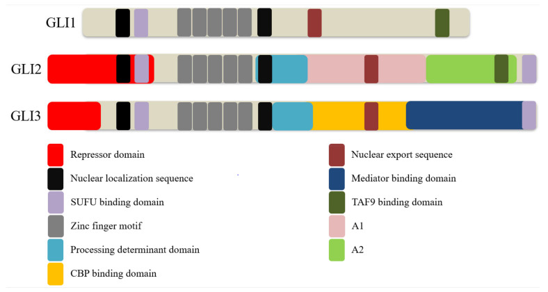
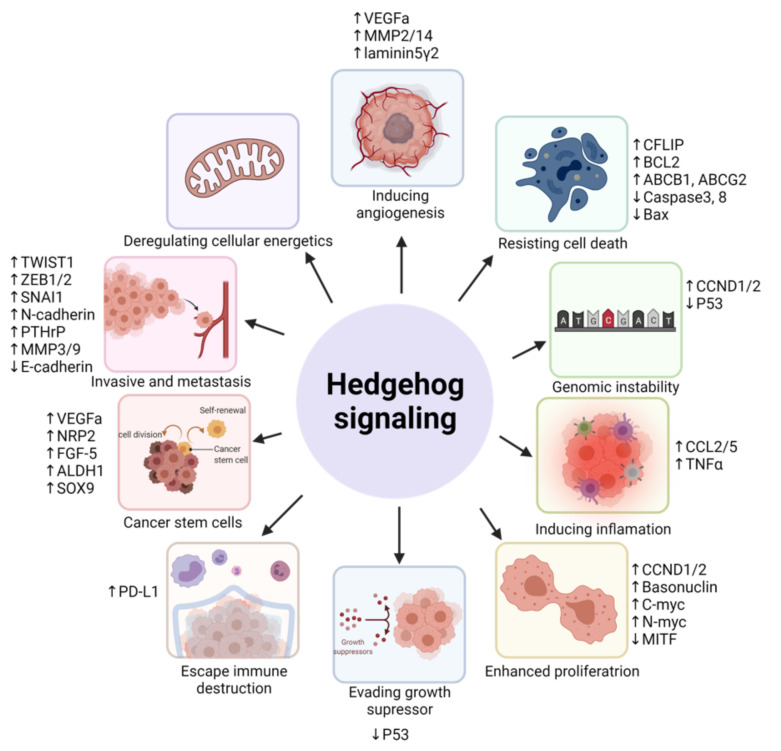
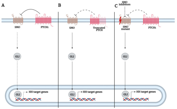
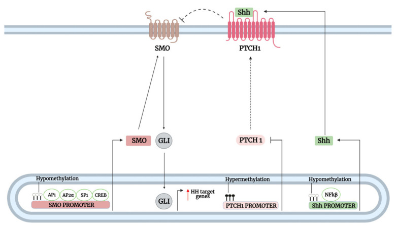
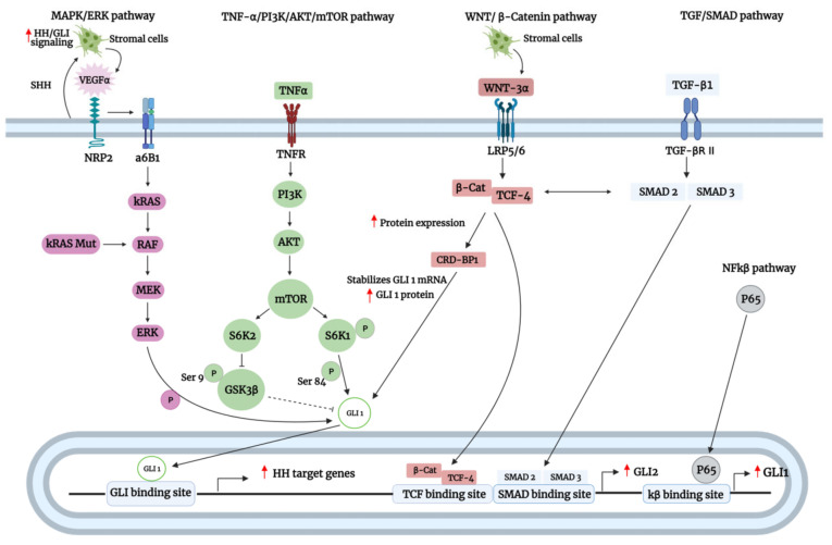
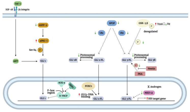
The Hedgehog (Hh)-glioma-associated oncogene homolog (GLI) signaling pathway is highly conserved among mammals, with crucial roles in regulating embryonic development as well as in cancer initiation and progression. The GLI transcription factors (GLI1, GLI2, and GLI3) are effectors of the Hh pathway and are regulated via Smoothened (SMO)-dependent and SMO-independent mechanisms. The SMO-dependent route involves the common Hh-PTCH-SMO axis, and mutations or transcriptional and epigenetic dysregulation at these levels lead to the constitutive activation of GLI transcription factors. Conversely, the SMO-independent route involves the SMO bypass regulation of GLI transcription factors by external signaling pathways and their interacting proteins or by epigenetic and transcriptional regulation of GLI transcription factors expression. Both routes of GLI activation, when dysregulated, have been heavily implicated in tumorigenesis of many known cancers, making them important targets for cancer treatment. Hence, this review describes the various SMO-dependent and SMO-independent routes of GLI regulation in the tumorigenesis of multiple cancers in order to provide a holistic view of the paradigms of hedgehog signaling networks involving GLI regulation. An in-depth understanding of the complex interplay between GLI and various signaling elements could help inspire new therapeutic breakthroughs for the treatment of Hh-GLI-dependent cancers in the future. Lastly, we have presented an up-to-date summary of the latest findings concerning the use of Hh inhibitors in clinical developmental studies and discussed the challenges, perspectives, and possible directions regarding the use of SMO/GLI inhibitors in clinical settings.

Keywords: GLI1 protein; cancer; clinical trial; epigenetic regulation; glioma-associated oncogene; hedgehog inhibitors; hedgehog pathway; mutations; noncanonical.


Conflict of interest statement

The authors declare no conflict of interest.

Figures

Figure 1
Figure 1
(A) The repression of Smoothened (SMO) by the Patched (PTCH) receptor in the absence of hedgehog (Hh) ligands promotes the interaction of Suppressor of Fused (SUFU) and glioma-associated oncogene homolog (GLI). G-protein coupled receptor 61 (GPR161) translocates to the primary cilium, which triggers high levels of cyclic adenosine monophosphate (CAMP). Elevated ciliary levels of CAMP maintain high levels of protein kinase A (PKA) activity, which phosphorylate GLI at P1-6 clusters. Consequently, phosphorylation of GLI by PKA prime its phosphorylation by casein kinase I (CKI) and glycogen synthase kinase 3 beta (GSK3β) further. Phosphorylated GLI is recognized by the β-TrCP, promoting its ubiquitination and partial proteasomal processing into a repressor. GLI repressor (GLIR) then translocates into the nucleus to repress target gene transcription. (B) The binding of the Hh ligand to the PTCH receptor alleviates its repression of SMO, allowing SMO translocation to the primary cilium. Activated SMO inhibits SUFU, allowing the dissociation of GLI from SUFU. Additionally, Gpr161 is removed from the primary cilium, causing low CAMP levels and PKA activity. The release of GLI from SUFU and low PKA activity results in the dephosphorylation of GLI, preventing its proteasomal processing into a repressor. Full-length GLI or GLI activator (GLIA) then translocates into the nucleus to transcribe target genes. Red upward triangle-headed arrow: upregulation; green downward triangle-headed arrow: downregulation; dotted black triangle-headed arrow: inactivation; bar-headed arrow: inhibition; dotted bar-headed arrow: loss of inhibition.
Figure 2
Figure 2
Schematic representation of the domains and motifs in glioma-associated oncogene homolog (GLI) proteins. All GLI proteins contain a well-conserved Supressor of Fused (SUFU)-binding domain, zinc finger motifs, nuclear localization sequences, and a nuclear export sequence. GLI2 and GLI3 contain both an N-terminal repressor and several C-terminal transactivation domains, unlike GLI1, which contains only a single transactivation domain reported so far. Additionally, GLI2 and GLI3 contain a second SUFU-binding domain at the C-terminal end critical for regulating nuclear GLI function. Both GLI2 and GLI3 contain a processing determinant domain that contributes to the proteolytic processing of these proteins into their repressor form with a more active role in GLI3 than GLI2. GLI2 contains two major transactivation domains, termed A1 and A2, while the GLI3 transactivation domain includes a CREB-binding protein (CBP)-binding domain and mediator-binding domain. Both GLI1 and GLI2 contain an α-helical herpes simplex viral protein 16-like activation domain that binds to TATA-box binding protein associated factor 9 (TAF9) due to the presence of a highly conserved FXXΦΦ (F = phenylalanine; X = any residue; Φ = any hydrophobic residue) motif in the domain. The FXXΦΦ motif is also conserved in GLI3 but does not bind to TAF9.
Figure 3
Figure 3
A simplified diagram on the role of Hh signaling activation in driving cancer hallmarks. Upward triangle-headed arrow: upregulation; downward triangle-headed arrow: downregulation.
Figure 4
Figure 4
A simplified illustration of Smoothened (SMO)-dependent glioma-associated oncogene homolog (GLI) regulation in the context of hedgehog (Hh) pathway mutations. (A) Under physiological conditions in adult tissues, Patched1 (PTCH1) functions by inhibiting SMO, which represses GLI function and prevents its translocation into the nucleus to activate target genes transcriptionally. (B) However, in cancer cells, loss of heterozygosity (LOH) of PTCH1 alleles results in the formation of a nonfunctional truncated PTCH1 protein. Consequently, this results in the constitutive activation of SMO, which promotes GLI activation and its translocation into the nucleus to activate Hh target genes transcriptionally. (C) Alternatively, constitutively active SMO M1/M2 mutants resistant to PTCH1 inhibition or inhibition by SMO inhibitors promote the sustained activation of GLI and its subsequent translocation into the nucleus to transcriptionally activate Hh target genes. Bar-headed arrow: inhibition; dotted bar-headed arrow: loss of inhibition; triangle-headed arrow: activation; dotted triangle-headed arrow: inactivation.
Figure 5
Figure 5
A simplified illustration of SMO-dependent GLI regulation in the context of transcriptional regulation. The binding of transcription factors AP1, AP2α, SP1, and CREB to promoter regions of SMO kickstarts the onset of its transcription. Similarly, the binding of NFκB to the NFκB binding site located within the Shh promoter induced the transcriptional upregulation of Shh. Furthermore, the increased transcriptional output of SMO and Shh can also occur as epigenetic events through hypomethylation of CpG islands that reside within promoters, which further promotes the binding of transcriptional machinery to promoters. Conversely, hypermethylation of the PTCH1 promoter leads to decreased transcriptional expression and consequently decreased production of PTCH1 protein. Together, these molecular events work in concert to further enhanced Hh pathway activity and promote tumorigenesis. Dotted triangle-headed arrow: inactivated function; dotted bar-headed arrow: loss of inhibition; red upward triangle-headed arrow: upregulation.
Figure 6
Figure 6
A schematic representation of the Smoothened (SMO)-independent regulation of glioma-associated oncogene homolog (GLI) transcription factors by oncogenic pathways. As shown above, GLI transcription factors can be regulated at the protein or transcriptional level depending on the oncogenic pathway involved. In the mitogen-activated protein kinase (MAPK)/ extracellular-signal-regulated kinase (ERK) pathway, sonic hedgehog (Shh) produced by tumor cells activates hedgehog (Hh)/GLI signaling in the stromal cells, leading to the upregulation of vascular endothelial growth factor A (VEGFa). Paracrine feedback of VEGFa to tumor cells is initiated upon binding of the VEGFa to neuropilin 2 (NRP2), which induces α6β1 integrin-mediated activation of kirsten rat sarcoma 2 viral oncogene homolog (KRAS)/ mitogen-activated protein kinase kinase (MEK)/ERK cascade. Active ERK1 then phosphorylates GLI1 protein, leading to its activation. Oncogenic KRAS mutations also lead to the constitutive activation of the MAPK/MEK/ERK pathway, consequently promoting GLI1 phosphorylation and activation. In the phosphoinositide 3-kinase (PI3K)/protein kinase B (AKT)/mechanistic target of rapamycin kinase (mTOR)pathway, tumor necrosis factor-alpha (TNFα) stimulation results in the activation of the mTOR complex, which in turn activates S6K2. Consequently, activated S6K2 phosphorylates glycogen synthase kinase 3 beta (GSK3β) at serine 9, leading to its inactivation. Inactivated GSK3β is not able to phosphorylate GLI1, relieving the inhibition of GSK3β on GLI1. Activation of the mTOR complex also activates S6K1 by phosphorylation, and activated S6K1, in turn, phosphorylate GLI1 at Ser9 to promote its activation. In the Wnt/β-catenin pathway, stromal cells produced Wnt3a that binds to the LRP5/6 receptor. The signal is then transduced to β-catenin, which forms a complex with T-cell factor 4 (TCF-4). The β-catenin-TCF-4 complex upregulates the protein expression of coding region determinant binding protein (CRD-BP), which stabilizes GLI1 mRNA and consequently enhances GLI1 protein levels. In the transforming growth factor-β (TGF-B)/SMAD pathway, stimulation by TGF-β results in the activation of SMAD2/3. SMAD2/3 cooperates with the β-catenin-TCF-4 complex to upregulate the expression of GLI2 by binding to the SMAD and TCF binding site within the GLI2 promoter. In the nuclear factor kappa B (NFκB) pathway, the p65 subunit of the NFκB complex binds to the kB binding site within the GLI1 promoter to initiate its transcription. Red upward triangle-headed arrow: upregulation.
Figure 7
Figure 7
A schematic representation of the Smoothened (SMO)-independent regulation of glioma-associated oncogene homolog (GLI) transcription factors by their interacting proteins. Apoptosis-stimulating of p53 protein 2 (ASPP2) deficiency enhanced the binding of atypical Protein Kinase C ι (aPKC-ι) with GLI1, which allows aPKC-ι to phosphorylate GLI1 at Ser84. The phosphorylated GLI1 is, in turn, activated, promoting its translocation into the nucleus to transcribe target genes. Galectin-1 (Gal-1) binds to β1 integrin to promote GLI1 activation. Mechanistically, activated β1 integrin forms a complex with insulin-like growth factor 1 receptor (IGF-1R) to promote protein kinase B (AKT) activation, leading to an increase in GLI1 activity. In the nucleus, SOX9 binds to the F-box region of β-TrCP, interfering with its binding to SKP1. The binding of SOX9 to β-TrCP tethers it within the nucleus, thus protecting GLI1 from degradation. Speckle-type POZ protein (SPOP) downregulation results in decreased ubiquitination of full-length GLI2/3 proteins, favoring their activation and nuclear translocation over their proteasomal processing into repressors. In the nucleus, the N-terminal domain (aa 1-68) of transcription factor forkhead box C1 (FOXC1) binds to the internal region (aa 898-1168) of GLI2, enhancing its DNA-binding and transcriptional-activating ability. The imbalance between Tyr216 and Ser9 phosphorylation of glycogen synthase kinase 3 beta (GSK3β) leads to its dysregulated function, thereby impairing its ability to phosphorylate full-length GLI3 proteins. Unphosphorylated full-length GLI3 proteins are not subjected to proteasomal processing into their repressors, allowing their translocation into the nucleus to transcribe target genes. Under androgen-deprived conditions, the downregulation of MED12 relieves its constraint on the full-length GLI3 proteins, resulting in their hyperactivation. Green downward triangle-headed arrow: downregulation; red upward triangle-headed arrow: upregulation; dotted bar-headed arrow: loss of inhibition.

References

    1. Nüsslein-volhard C., Wieschaus E. Mutations Affecting Segment Number and Polarity in Drosophila. Nature. 1980;287:795–801. doi: 10.1038/287795a0. - DOI - PubMed
    1. Carballo G.B., Honorato J.R., De Lopes G.P.F., de Sampaio E., Spohr T.C.L. A Highlight on Sonic Hedgehog Pathway. Cell Commun. Signal. 2018;16:11. doi: 10.1186/s12964-018-0220-7. - DOI - PMC - PubMed
    1. Mastronardi F.G., Dimitroulakos J., Kamel-Reid S., Manoukian A.S. Co-Localization of Patched and Activated Sonic Hedgehog to Lysosomes in Neurons. Neuroreport. 2000;11:581–585. doi: 10.1097/00001756-200002280-00030. - DOI - PubMed
    1. Rohatgi R., Milenkovic L., Corcoran R.B., Scott M.P. Hedgehog Signal Transduction by Smoothened: Pharmacologic Evidence for a 2-Step Activation Process. Proc. Natl. Acad. Sci. USA. 2009;106:3196–3201. doi: 10.1073/pnas.0813373106. - DOI - PMC - PubMed
    1. Hsu S.H.C., Zhang X., Yu C., Li Z.J., Wunder J.S., Hui C.C., Alman B.A. Kif7 Promotes Hedgehog Signaling in Growth Plate Chondrocytes by Restricting the Inhibitory Function of Sufu. Development. 2011;138:3791–3801. doi: 10.1242/dev.069492. - DOI - PubMed

LinkOut - more resources