Review
. 2021 Sep 18;9(9):1265.
doi: 10.3390/biomedicines9091265.

Stationed or Relocating: The Seesawing EMT/MET Determinants from Embryonic Development to Cancer Metastasis

Affiliations
Review

Stationed or Relocating: The Seesawing EMT/MET Determinants from Embryonic Development to Cancer Metastasis

Chien-Hsiu Li et al. Biomedicines. .

Abstract

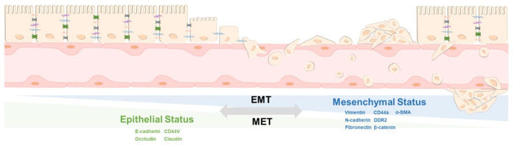
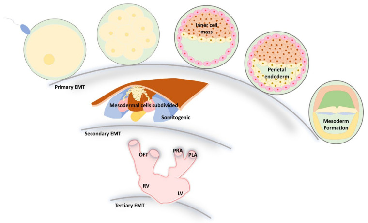
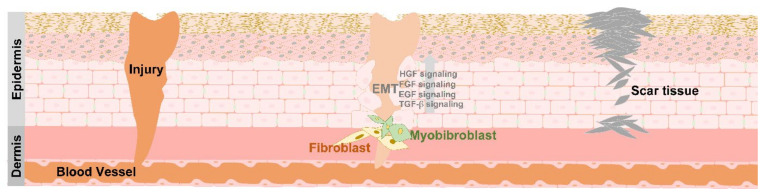
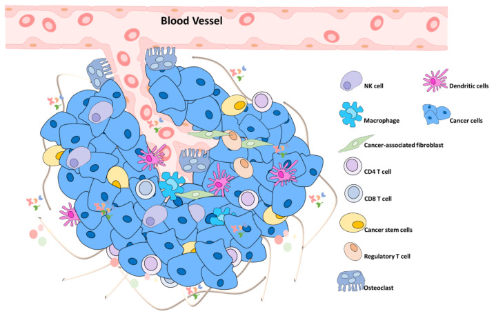
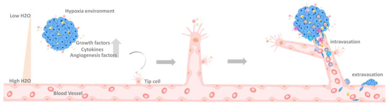
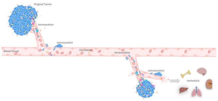
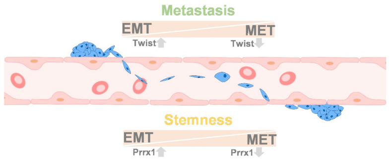
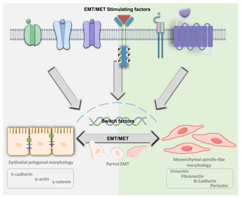
Epithelial and mesenchymal transition mechanisms continue to occur during the cell cycle and throughout human development from the embryo stage to death. In embryo development, epithelial-mesenchymal transition (EMT) can be divided into three essential steps. First, endoderm, mesoderm, and neural crest cells form, then the cells are subdivided, and finally, cardiac valve formation occurs. After the embryonic period, the human body will be subjected to ongoing mechanical stress or injury. The formation of a wound requires EMT to recruit fibroblasts to generate granulation tissues, repair the wound and re-create an intact skin barrier. However, once cells transform into a malignant tumor, the tumor cells acquire the characteristic of immortality. Local cell growth with no growth inhibition creates a solid tumor. If the tumor cannot obtain enough nutrition in situ, the tumor cells will undergo EMT and invade the basal membrane of nearby blood vessels. The tumor cells are transported through the bloodstream to secondary sites and then begin to form colonies and undergo reverse EMT, the so-called "mesenchymal-epithelial transition (MET)." This dynamic change involves cell morphology, environmental conditions, and external stimuli. Therefore, in this manuscript, the similarities and differences between EMT and MET will be dissected from embryonic development to the stage of cancer metastasis.

Keywords: EMT; MET; embryonic; tissue repair; tumorigenesis.


Conflict of interest statement

The authors declare no conflict of interest.

Figures

Figure 1
Figure 1
The regulation of EMT and MET. Epithelial cells can transform the original cell type from epithelial status to mesenchymal status through the EMT process, and its ability to move, such as entering the circulation system and then returning to epithelial status, through the conversion of MET. Therefore, the level of EMT and MET mobility and its corresponding markers to distinguish the types of EMT and MET is described.
Figure 2
Figure 2
EMT progression of development in the first, secondary, and tertiary embryo stages. EMT plays an essential role in embryonic development and is closely related to the movement of different germ layers to specific locations for further differentiation. During embryogenesis, the embryo can undergo three major EMT processes. First, once the zygote is formed, the cells will move to a specific location to distribute three primary germ layers, endoderm, mesoderm, and ectoderm, called primary EMT. The second EMT can be observed in the epithelial structure among the mesoderm, notochord, neural tube, and somite, which undergo processes such as endocrine cell formation. Finally, the formation of mesenchymal cells, such as cardiac cushions as cardiac valve precursors, is the most appropriate model to describe tertiary EMT. OFT  =  Outflow Tract; RV  =  Right Ventricle; PRA  =  Primitive Right Atrium; PLA  =  Primitive Left Atrium; LV  =  Left Ventricle.
Figure 3
Figure 3
Wound healing progression. The diagram shows wound healing progression. Different signal transmissions interact with neighboring cells through EMT when the tissue is damaged to repair the wound. The related signaling, such as FGF, EGF, TGF-b, and HGF, can induce cell migration by redefined epithelial, mesenchymal, and secret matrix metalloproteinase to remodel extracellular components. In addition, cells, such as Keratinocyte and fibroblast, can undergo the EMT and cellular proliferation process to participate in wound healing-related processes.
Figure 4
Figure 4
Primary tumor. The tumor microenvironment contains tumor stem cells and tumor-associated extracellular components, such as immune cells. Mainly, cancer cells, such as tumor-associated macrophages, tumor-associated adipocytes, tumor endothelial cells, and tumor-associated fibroblast, can change the characteristics of neighboring cells in various ways and further interact with each other to increase the degree of tumor heterogeneity.
Figure 5
Figure 5
The progression of extravasation. Cancer cells can migrate from carcinoma in situ through blood vessels to other parts of the body through the extravasation process. Under this process, the hypoxia environment can stimulate tumor cells to secret chemotactic factors, such as growth factors, cytokines, and angiogenesis-related factors, stimulating epithelial cells for blood vessel interaction through angiogenesis. In such cases, tumor cells will develop intravasation into the blood vessel and the extravasation process to uptake more nutrients. Up arrow means upregulation.
Figure 6
Figure 6
Metastasis progression. The illustration shows how cancer cells obtain more nutrients through angiogenesis and grow outwards. Through extravasation, they can transfer to other parts of the body for further expansion. The original tumor cells can undergo EMT and intravasation and circulate in blood vessels. Under the EMT process, cells survive in strict circumstances and through extravasation metastasis to distinct tissues, such as the brain, kidney, lung, liver, and bone.
Figure 7
Figure 7
The theory of cancer stem cells. Changes in Twist and Prrx1 between EMT and MET explain the possible conversions between cell type and function. When overexpressed tumor cells twist, cells with more EMT phenotypes and undergo cell motility; once cells metastasize to the proper location, the related twist will be downregulated, and the MET process enables cell colonization. However, such a process was distinct to the cell’s stemness ability. Instead, the expression of Prxx1 could be activated by EMT but suppressed stemness. Moreover, the MET process ability stemness activity increased in colonization, in which Prxx1 was downregulated. Up arrow means upregulation. Down arrow means downregulation.
Figure 8
Figure 8
Simulated molecular network between EMT and MET. The simulation of molecular interactions between EMT or MET hallmarks and the related biological functions of participation was conducted through prediction methods, in which gene ontology is linked to cytoskeleton remodeling, metastasis, remodeling the microenvironment, and embryogenesis-related process. (MET- or EMT-related molecules were downloaded from GSEA datasets, and the analyzed interaction map results were output from the Ingenuity Pathway Analysis.).
Figure 8
Figure 8
Simulated molecular network between EMT and MET. The simulation of molecular interactions between EMT or MET hallmarks and the related biological functions of participation was conducted through prediction methods, in which gene ontology is linked to cytoskeleton remodeling, metastasis, remodeling the microenvironment, and embryogenesis-related process. (MET- or EMT-related molecules were downloaded from GSEA datasets, and the analyzed interaction map results were output from the Ingenuity Pathway Analysis.).
Figure 9
Figure 9
The upstream regulators of EMT/MET dominate both embryonic development and cancer progression. A simulation of molecular interactions of EMT and MET hallmarks upstream regulators and the related biological functions of participation was conducted through prediction methods.
Figure 9
Figure 9
The upstream regulators of EMT/MET dominate both embryonic development and cancer progression. A simulation of molecular interactions of EMT and MET hallmarks upstream regulators and the related biological functions of participation was conducted through prediction methods.
Figure 10
Figure 10
The molecular mechanism of EMT/MET. To date, multiple essential embryogenesis factors have been proven to contribute to EMT/MET by control diverse switch factor activity, in which the epithelial or mesenchymal status has been well characterized, but among them, the intermediated status, partial EMT empower cancer stemness, drug-resistant, and collective motility, such plasticity contribute to tumor heterogeneity of tumors.

Similar articles

Cited by

References

    1. Thiery J.P., Sleeman J.P. Complex networks orchestrate epithelial-mesenchymal transitions. Nat. Rev. Mol. Cell. Biol. 2006;7:131–142. doi: 10.1038/nrm1835. - DOI - PubMed
    1. Thomson J.A., Itskovitz-Eldor J., Shapiro S.S., Waknitz M.A., Swiergiel J.J., Marshall V.S., Jones J.M. Embryonic stem cell lines derived from human blastocysts. Science. 1998;282:1145–1147. doi: 10.1126/science.282.5391.1145. - DOI - PubMed
    1. Li Q., Hutchins A.P., Chen Y., Li S., Shan Y., Liao B., Zheng D., Shi X., Li Y., Chan W.Y., et al. A sequential EMT-MET mechanism drives the differentiation of human embryonic stem cells towards hepatocytes. Nat. Commun. 2017;8:1–12. doi: 10.1038/ncomms15166. - DOI - PMC - PubMed
    1. Massri A.J., Schiebinger G.R., Berrio A., Wang L., Wray G.A., McClay D.R. Methodologies for Following EMT In Vivo at Single Cell Resolution. Methods Mol. Biol. 2021;2179:303–314. doi: 10.1007/978-1-0716-0779-4_23. - DOI - PMC - PubMed
    1. Osborne C.C., Perry K.J., Shankland M., Henry J.Q. Ectomesoderm and epithelial-mesenchymal transition-related genes in spiralian development. Dev. Dyn. 2018;247:1097–1120. doi: 10.1002/dvdy.24667. - DOI - PubMed

LinkOut - more resources