. 2021 Aug 25;11(9):1273.
doi: 10.3390/biom11091273.

Mutations of SARS-CoV-2 RBD May Alter Its Molecular Structure to Improve Its Infection Efficiency

Affiliations

Mutations of SARS-CoV-2 RBD May Alter Its Molecular Structure to Improve Its Infection Efficiency

Ahmed L Alaofi et al. Biomolecules. .

Abstract

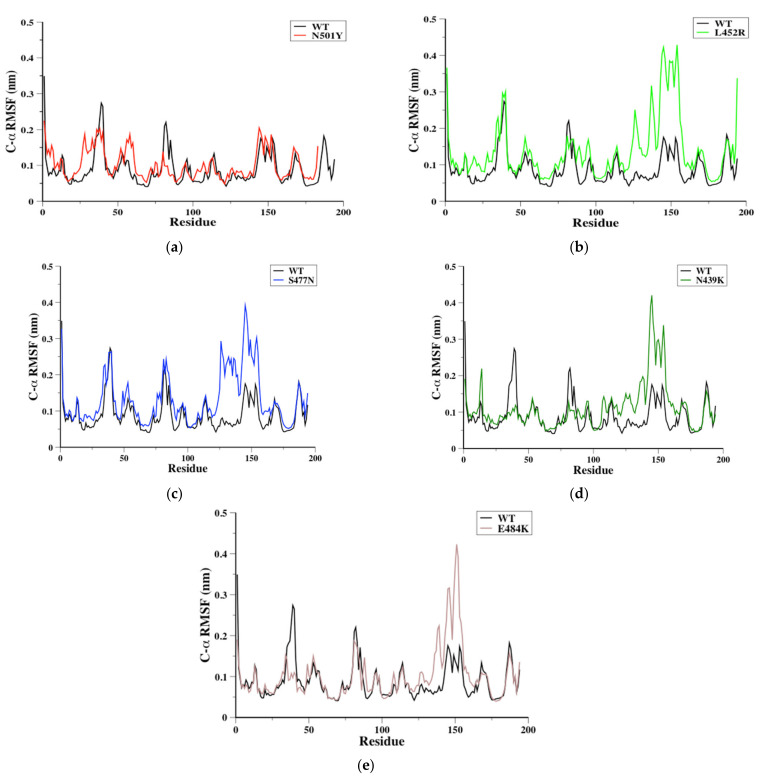
The receptor-binding domain (RBD) of severe acute respiratory syndrome coronavirus 2 (SARS-CoV-2) mediates the viral-host interaction and is a target for most neutralizing antibodies. Nevertheless, SARS-CoV-2 RBD mutations pose a threat due to their role in host cell entry via the human angiotensin-converting enzyme 2 receptor that might strengthen SARS-CoV-2 infectivity, viral load, or resistance against neutralizing antibodies. To understand the molecular structural link between RBD mutations and infectivity, the top five mutant RBDs (i.e., N501Y, E484K L452R, S477N, and N439K) were selected based on their recorded case numbers. These mutants along with wild-type (WT) RBD were studied through all-atom molecular dynamics (MD) simulations of 100 ns. The principal component analysis and the free energy landscape were used too. Interestingly, N501Y, N439K, and E484K mutations were observed to increase the rigidity in some RBD regions while increasing the flexibility of the receptor-binding motif (RBM) region, suggesting a compensation of the entropy penalty. However, S477N and L452R RBDs were observed to increase the flexibility of the RBM region while maintaining similar flexibility in other RBD regions in comparison to WT RBD. Therefore, both mutations (especially S477N) might destabilize the RBD structure, as loose conformation compactness was observed. The destabilizing effect of S477N RBD was consistent with previous work on S477N mutation. Finally, the free energy landscape results showed that mutations changed WT RBD conformation while local minima were maintained for all mutant RBDs. In conclusion, RBD mutations definitely impact the WT RBD structure and conformation as well as increase the binding affinity to angiotensin-converting enzyme receptor.

Keywords: RBD flexibility; SARS-CoV-2; free energy landscape; molecular dynamics simulations; mutant RBDs; principle component analysis; wild-type RBD.


Conflict of interest statement

The authors declare no conflict of interest.

Figures

Figure 1
Figure 1
The C-α root mean square deviation (C-α RMSD) in nm was depicted for wild-type (WT) (black), N501Y (red), L452R (light green), S477N (blue), N439K (green), and E484K (cyan) RBDs during the 100-ns MD simulations.
Figure 2
Figure 2
The C-α root mean square fluctuation (C-α RMSF) in nm for WT RBD aligned with either N501Y (a), L452R (c), S477N (d), N439K (e), or E484K (f) RBDs as a function of RBD residues obtained from the 100-ns MD simulations. The WT RBD cartoon representative (b) was plotted to indicate the RBD regions of interest.
Figure 3
Figure 3
To illustrate RBD region, the ribbon representative of 100 ns conformer of WT RBD is depicted with red color and RBM is colored in blue and black arrows indicate RBM loop regions (a). Ten conformers obtained every 10 ns were aligned together from each MD simulations system. The ribbon representative for WT, N501Y, L452R, S477N, N439K, or E484K RBDs was aligned together in order to observe the conformational changes of RBD regions during the simulations (b). A cartoon representative was plotted to show the hydrogen bonds formed by the N501Y and L452R mutations; small boxes provide enlarged images of the hydrogen bonds formed for N501Y (green) and L452R (cyan) RBDs (c).
Figure 4
Figure 4
The radius of gyration (Rg) in nm was plotted versus simulation time (ps). WT (black), N501Y (red), N439K (green), and E484K (cyan) RBDs showed a relatively similar Rg for all of the simulations (a). L452R (light green) and S477N (blue) were different from WT RBD over most of the 100-ns simulations. However, L452R showed similar conformations in the last frame of MD simulations.
Figure 5
Figure 5
Projection of the motion of WT RBD (black color) aligned with either (a) N501Y (red color), (b) L452R (light green color), (c) S477N (blue color), (d) N439K (green color), or (e) E484K (cyan color) mutant receptor-binding domains (RBDs) along with the first two principal eigenvectors in nm.
Figure 6
Figure 6
Porcupine plots showing the motion across the first principal component (PC) in WT RBD (a) and N501Y (b), L452R (c), S477N (d), N439K (e), and E484K (f) mutants. The arrows reflect the direction of the correlated motion and the extent of the motion.
Figure 7
Figure 7
The free energy landscape (FEL) was obtained during the 100-ns MD simulations for each RBD system: WT RBD (a), N501Y (b), L452R (c), S477N (d), N439K (e), or E484K (f) RBDs.
Figure 7
Figure 7
The free energy landscape (FEL) was obtained during the 100-ns MD simulations for each RBD system: WT RBD (a), N501Y (b), L452R (c), S477N (d), N439K (e), or E484K (f) RBDs.

Similar articles

Cited by

References

    1. WHO Coronavirus (COVID-19) Dashboard. [(accessed on 6 June 2021)]. Available online: https://covid19.who.int.
    1. Shang J., Wan Y., Luo C., Ye G., Geng Q., Auerbach A., Li F. Cell Entry Mechanisms of SARS-CoV-2. Proc. Natl. Acad. Sci. USA. 2020;117:11727–11734. doi: 10.1073/pnas.2003138117. - DOI - PMC - PubMed
    1. Gao H., Yao H., Yang S., Li L. From SARS to MERS: Evidence and Speculation. Front. Med. 2016;10:377–382. doi: 10.1007/s11684-016-0466-7. - DOI - PMC - PubMed
    1. Walls A.C., Park Y.-J., Tortorici M.A., Wall A., McGuire A.T., Veesler D. Structure, Function, and Antigenicity of the SARS-CoV-2 Spike Glycoprotein. Cell. 2020;181:281–292. doi: 10.1016/j.cell.2020.02.058. - DOI - PMC - PubMed
    1. Ye Z.-W., Yuan S., Yuen K.-S., Fung S.-Y., Chan C.-P., Jin D.-Y. Zoonotic Origins of Human Coronaviruses. Int. J. Biol. Sci. 2020;16:1686–1697. doi: 10.7150/ijbs.45472. - DOI - PMC - PubMed

Publication types

MeSH terms

Substances