Review
. 2021 Aug 27;11(9):1284.
doi: 10.3390/biom11091284.

Impact of G-Quadruplexes on the Regulation of Genome Integrity, DNA Damage and Repair

Affiliations
Review

Impact of G-Quadruplexes on the Regulation of Genome Integrity, DNA Damage and Repair

Anzhela V Pavlova et al. Biomolecules. .

Abstract

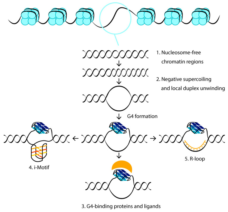
DNA G-quadruplexes (G4s) are known to be an integral part of the complex regulatory systems in both normal and pathological cells. At the same time, the ability of G4s to impede DNA replication plays a critical role in genome integrity. This review summarizes the results of recent studies of G4-mediated genomic and epigenomic instability, together with associated DNA damage and repair processes. Although the underlying mechanisms remain to be elucidated, it is known that, among the proteins that recognize G4 structures, many are linked to DNA repair. We analyzed the possible role of G4s in promoting double-strand DNA breaks, one of the most deleterious DNA lesions, and their repair via error-prone mechanisms. The patterns of G4 damage, with a focus on the introduction of oxidative guanine lesions, as well as their removal from G4 structures by canonical repair pathways, were also discussed together with the effects of G4s on the repair machinery. According to recent findings, there must be a delicate balance between G4-induced genome instability and G4-promoted repair processes. A broad overview of the factors that modulate the stability of G4 structures in vitro and in vivo is also provided here.

Keywords: DNA damage; DNA repair; G-quadruplex; G-quadruplex-binding proteins; genomic instability.


Conflict of interest statement

The authors declare no conflict of interest.

Figures

Figure 1
Figure 1
Cellular factors contributing to the folding and stabilization of G4 structures. Blue circles represent histones, G4-binding proteins and low molecular weight ligands are shown in yellow.
Figure 2
Figure 2
Scheme of G4-mediated genesis of DSBs after the second round of replication and subsequent DSB repair by the TMEJ mechanism with the formation of typical mutational patterns. Hybridization based on microhomology (in orange) and DNA synthesis by DNA Pol θ results in the deletion of sites external to microhomology (dashed line). Switching the template to another microhomologous sequence (in blue) leads to a Pol θ-mediated sequence insertion adjacent to the microhomology (in red). Several template switches can result in more reinsertions.
Figure 3
Figure 3
The structure of the complex between part of the DHX36 helicase (RSM peptide, in pink and green) and parallel G4 with heterocyclic bases marked in blue (PDB 2N21 [90]). Three positively charged amino acid residues (K8, R10 and K19, circled) form electrostatic interactions with phosphate groups of G4. The amino acids of the α-helix stack with the external G-tetrad, while the AKKQ motif (in green) anchors the peptide in the G4 groove.
Figure 4
Figure 4
(a) Positions in G4s of different topologies that are most susceptible to oxidative damage. (b) Replacement of damaged guanine in the G4 core with available dG in loops or spare G-tracts helps to maintain the G4 structure and increases the accessibility of damaged dG residues for repair proteins.
Figure 5
Figure 5
The relative preference of BER glycosylases toward oxidative guanine lesions in different structural contexts (various G4 topologies (left) versus single- and double-stranded DNAs (right)) as substrates for repair. The type of damage is shown above the arrows, the thickness of which reflects the repair efficiency: the thicker lines represent the better substrate, the thinner lines designate the relatively weaker enzyme activation, while the crossed arrows indicate the lack of the corresponding BER glycosylase activity against a specific oxidative lesion in a definite context.
Figure 6
Figure 6
(a) Induction of BER-dependent G4 formation by oxidative stress. Removal of 8-oxoG by OGG1 glycosylase results in an AP site that destabilizes the DNA duplex and makes the formation of G4 by a G4 motif with five G-tracts more favorable. (b) Recognition, stabilization and protection against G4-unwinding helicases of G4 structures by the Zuo1 protein stimulates the repair of UV-induced damage (T=T corresponds to thymine dimer) via the NER pathway by recruiting the Rad4/23 complex.
Figure 7
Figure 7
Efficiency of ecMMR protein-induced hydrolysis of linear DNA duplexes with an embedded parallel G4 and a MutH recognition site, which differ in the mismatch position. (a) DNA models used in this work; their names are shown on the left and right, and the sequences are the same as in [35]. 5′-Gm6ATC-3′/3′-CTAG-5′ corresponds to MutH recognition site, and black arrows indicate the position of DNA cleavage by the MutH endonuclease. Pink asterisks represent the TAMRA fluorophore at the 3′-end of the unmethylated daughter strand. 3′-Labeled cleavage products were separated from intact DNA strands by gel electrophoresis under denaturing conditions (7 M urea), allowing for evaluation of nicking potency; for the experimental conditions, see [35]. (b) Efficiency of DNA hydrolysis induced by ecMMR proteins (p < 0.05). Data were obtained for the MutH alone (250 nM concentration) and for the combinations of MutH with 250 nM MutS or 250 nM MutL, as well as with both 250 nM ecMutS and 250 nM ecMutL (all protein concentrations were calculated per monomer). The reaction mixtures were incubated at 37 °C for 1 h. These results have not yet been published.

Similar articles

Cited by

References

    1. Marušič M., Šket P., Bauer L., Viglasky V., Plavec J. Solution-state structure of an intramolecular G-quadruplex with propeller, diagonal and edgewise loops. Nucleic Acids Res. 2012;40:6946–6956. doi: 10.1093/nar/gks329. - DOI - PMC - PubMed
    1. Dolinnaya N.G., Ogloblina A.M., Yakubovskaya M.G. Structure, properties, and biological relevance of the DNA and RNA G-quadruplexes: Overview 50 years after their discovery. Biochemistry. 2016;81:1602–1649. doi: 10.1134/S0006297916130034. - DOI - PMC - PubMed
    1. Fujii T., Podbevšek P., Plavec J., Sugimoto N. Effects of metal ions and cosolutes on G-quadruplex topology. J. Inorg. Biochem. 2017;166:190–198. doi: 10.1016/j.jinorgbio.2016.09.001. - DOI - PubMed
    1. Miyoshi D. Structural transition from antiparallel to parallel G-quadruplex of d(G4T4G4) induced by Ca2+ Nucleic Acids Res. 2003;31:1156–1163. doi: 10.1093/nar/gkg211. - DOI - PMC - PubMed
    1. Chung W.J., Heddi B., Schmitt E., Lim K.W., Mechulam Y., Phan A.T. Structure of a left-handed DNA G-quadruplex. Proc. Natl. Acad. Sci. USA. 2015;112:2729–2733. doi: 10.1073/pnas.1418718112. - DOI - PMC - PubMed

Publication types

LinkOut - more resources