Review
. 2021 Sep 21;11(9):1389.
doi: 10.3390/biom11091389.

Mechanosensitivity in Pulmonary Circulation: Pathophysiological Relevance of Stretch-Activated Channels in Pulmonary Hypertension

Affiliations
Review

Mechanosensitivity in Pulmonary Circulation: Pathophysiological Relevance of Stretch-Activated Channels in Pulmonary Hypertension

Solène Barbeau et al. Biomolecules. .

Abstract

A variety of cell types in pulmonary arteries (endothelial cells, fibroblasts, and smooth muscle cells) are continuously exposed to mechanical stimulations such as shear stress and pulsatile blood pressure, which are altered under conditions of pulmonary hypertension (PH). Most functions of such vascular cells (e.g., contraction, migration, proliferation, production of extracellular matrix proteins, etc.) depend on a key event, i.e., the increase in intracellular calcium concentration ([Ca2+]i) which results from an influx of extracellular Ca2+ and/or a release of intracellular stored Ca2+. Calcium entry from the extracellular space is a major step in the elevation of [Ca2+]i, involving a variety of plasmalemmal Ca2+ channels including the superfamily of stretch-activated channels (SAC). A common characteristic of SAC is that their gating depends on membrane stretch. In general, SAC are non-selective Ca2+-permeable cation channels, including proteins of the TRP (Transient Receptor Potential) and Piezo channel superfamily. As membrane mechano-transducers, SAC convert physical forces into biological signals and hence into a cell response. Consequently, SAC play a major role in pulmonary arterial calcium homeostasis and, thus, appear as potential novel drug targets for a better management of PH.

Keywords: Piezo channel; TRP channel; calcium; endothelial cell; fibroblast; mechanosensitive channel; pulmonary arterial smooth muscle cell; pulmonary artery; pulmonary hypertension; vascular cell.


Conflict of interest statement

The authors declare no conflict of interest.

Figures

Figure 1
Figure 1
Hemodynamic forces acting on the vessel wall. Section of an artery wall showing that the endothelial cells, forming the inner tunica, are longitudinally aligned, whereas smooth muscle cells, forming the median layer, are circumferentially aligned; the surrounding adventitia predominantly includes fibroblasts and matrix. Shear stress, frictional force generated by blood flow, is parallel to the vessel wall, whereas blood pressure is perpendicular to the vessel wall, causing circumferential and longitudinal stretching. Beside blood mechanical forces, composition of extracellular matrix, contributing to arterial stiffness, may itself modulate compliance and mechanotransduction in the vessel wall.
Figure 2
Figure 2
Activation mechanisms of SAC. Three general models are proposed: (a) In the “bilayer model”, the tension developed (red arrow) in the lipid bilayer itself is directly responsible for channel gating. (b) In the “tether model”, the force is transmitted to the channel via proteins located in the extracellular matrix, the cytoskeleton, or both. Tensions are conveyed by these accessory proteins to induce the channel opening. (c) In the “secondary signal model”, the channel activation depends on a distant mechanical-sensitive protein generating diffusible second messenger or channel phosphorylation.
Figure 3
Figure 3
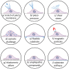
Experimental strategies to investigate SAC in cells. At the cellular level, several strategies can be used to activate SAC. The most commonly used are based on membrane deformation: (a) applying positive or negative pressure to the back end of the patch pipette, (b) poking of the cell membrane by a piezo-driven glass pipette, (c) modifying the perfusion flow or the viscosity of the solution, (d) using osmotic challenges: hypotonicity induces cell swelling, whilst hypertonicity evokes cell-shrinkage, (e) elongating thin elastic silicone membrane where cells are seeded, (f) applying magnetic field to specific ligands coated with magnetic particles on the cells, (g) seeding cells on elastomeric pillars to apply force to specific parts of the cells, and (h) using crenators and cup formers (amphipathic compounds) to induce crenation or cup shapes. (i) Another alternative consists in culturing cells in matrices of different stiffness, to evaluate the impact of the environment matrix and more especially its stiffness.
Figure 4
Figure 4
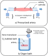
Experimental strategies to investigate SAC in vessels. At the tissue level, the effects of stretch can also be studied in whole vessels using (a) arteriography: the microvessel is cannulated at both ends with glass micropipettes and placed in a microvascular flow system chamber, allowing intraluminal pressure increase via modulation of inlet and outlet pressures (Pin and Pout, respectively); or (b) myography: one end of the segment is anchored to a stationary support and the other end is connected to a force-displacement transducer to monitor the vessel contraction under resting tension corresponding to an adapted transmural pressure.
Figure 5
Figure 5
Schematic view illustrating the multifunctional contribution of SAC in the pathogenesis of PH. Red arrows indicate PH-induced modifications of cellular processes in pulmonary arterial vascular cells.

References

    1. Yamashiro Y., Yanagisawa H. The Molecular Mechanism of Mechanotransduction in Vascular Homeostasis and Disease. Clin. Sci. 2020;134:2399–2418. doi: 10.1042/CS20190488. - DOI - PubMed
    1. Bayliss W.M. On the Local Reactions of the Arterial Wall to Changes of Internal Pressure. J. Physiol. 1902;28:220–231. doi: 10.1113/jphysiol.1902.sp000911. - DOI - PMC - PubMed
    1. Liu F., Haeger C.M., Dieffenbach P.B., Sicard D., Chrobak I., Coronata A.M., Suarez Velandia M.M., Vitali S., Colas R.A., Norris P.C., et al. Distal Vessel Stiffening Is an Early and Pivotal Mechanobiological Regulator of Vascular Remodeling and Pulmonary Hypertension. JCI Insight. 2016;1:e86987. doi: 10.1172/jci.insight.86987. - DOI - PMC - PubMed
    1. Galie N., Humbert M., Vachiery J.L., Gibbs S., Lang I., Torbicki A., Simonneau G., Peacock A., Vonk Noordegraaf A., Beghetti M., et al. 2015 ESC/ERS Guidelines for the Diagnosis and Treatment of Pulmonary Hypertension: The Joint Task Force for the Diagnosis and Treatment of Pulmonary Hypertension of the European Society of Cardiology (ESC) and the European Respiratory Society (ERS): Endorsed by: Association for European Paediatric and Congenital Cardiology (AEPC), International Society for Heart and Lung Transplantation (ISHLT) Eur. Heart J. 2016;37:67–119. doi: 10.1093/eurheartj/ehv317. - DOI - PubMed
    1. Humbert M., Galie N., McLaughlin V.V., Rubin L.J., Simonneau G. An Insider View on the World Symposium on Pulmonary Hypertension. Lancet Respir. Med. 2019;7:484–485. doi: 10.1016/S2213-2600(19)30111-0. - DOI - PubMed

Publication types

Substances