. 2021 Aug 25;10(9):1033.
doi: 10.3390/antibiotics10091033.

Antibiotics Used in Empiric Treatment of Ocular Infections Trigger the Bacterial Rcs Stress Response System Independent of Antibiotic Susceptibility

Affiliations

Antibiotics Used in Empiric Treatment of Ocular Infections Trigger the Bacterial Rcs Stress Response System Independent of Antibiotic Susceptibility

Nathaniel S Harshaw et al. Antibiotics (Basel). .

Abstract

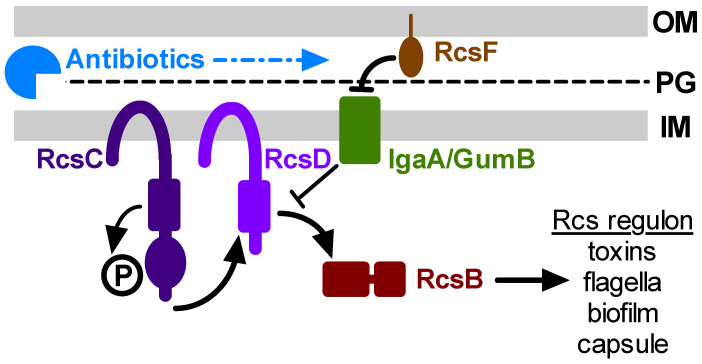
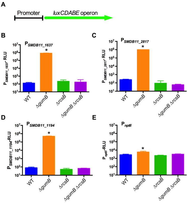
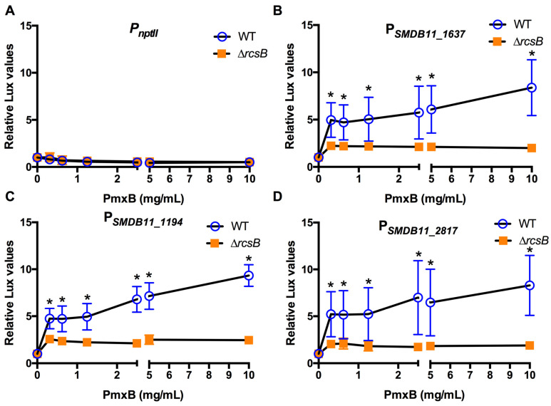
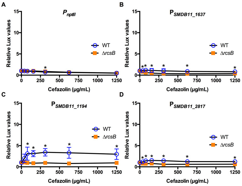
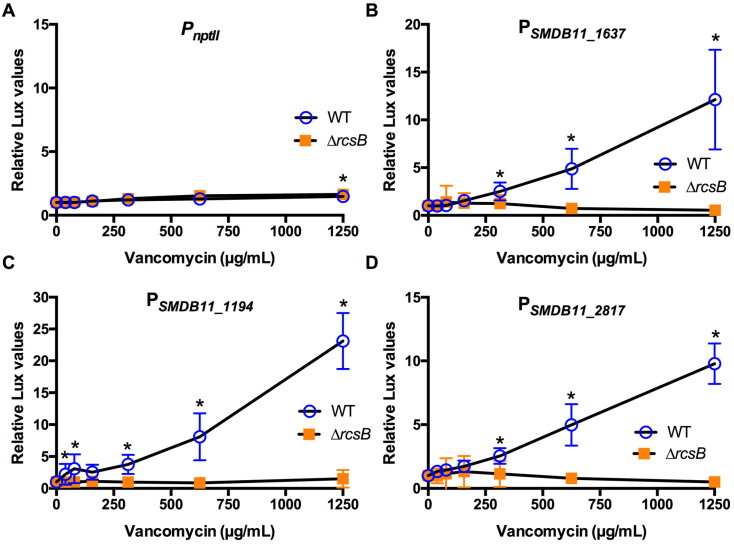
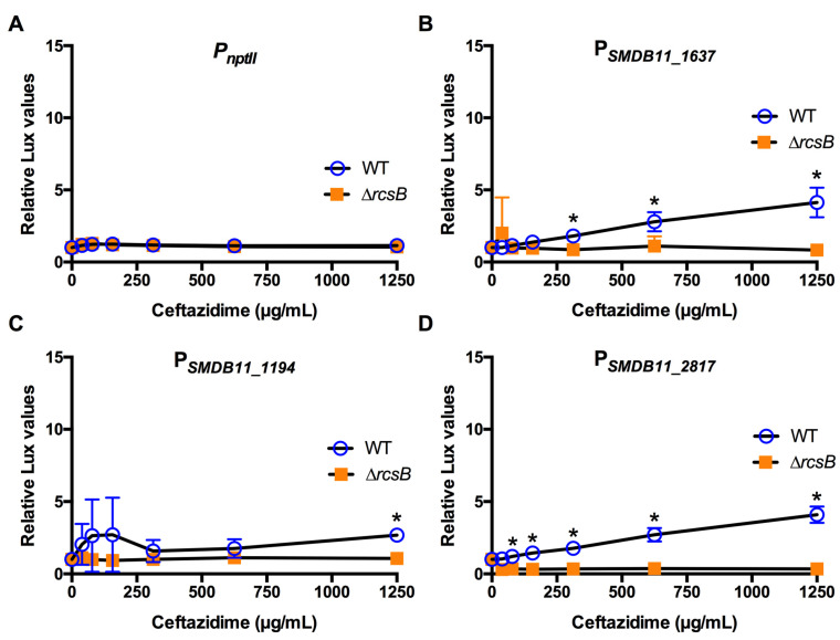
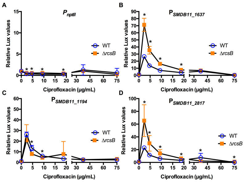
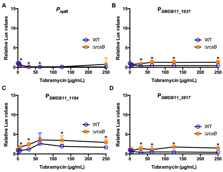
The Rcs phosphorelay is a bacterial stress response system that responds to envelope stresses and in turn controls several virulence-associated pathways, including capsule, flagella, and toxin biosynthesis, of numerous bacterial species. The Rcs system also affects antibiotic tolerance, biofilm formation, and horizontal gene transfer. The Rcs system of the ocular bacterial pathogen Serratia marcescens was recently demonstrated to influence ocular pathogenesis in a rabbit model of keratitis, with Rcs-defective mutants causing greater pathology and Rcs-activated strains demonstrating reduced inflammation. The Rcs system is activated by a variety of insults, including β-lactam antibiotics and polymyxin B. In this study, we developed three luminescence-based transcriptional reporters for Rcs system activity and used them to test whether antibiotics used for empiric treatment of ocular infections influence Rcs system activity in a keratitis isolate of S. marcescens. These included antibiotics to which the bacteria were susceptible and resistant. Results indicate that cefazolin, ceftazidime, polymyxin B, and vancomycin activate the Rcs system to varying degrees in an RcsB-dependent manner, whereas ciprofloxacin and tobramycin activated the promoter fusions, but in an Rcs-independent manner. Although minimum inhibitory concentration (MIC) analysis demonstrated resistance of the test bacteria to polymyxin B and vancomycin, the Rcs system was activated by sub-inhibitory concentrations of these antibiotics. Together, these data indicate that a bacterial stress system that influences numerous pathogenic phenotypes and drug-tolerance is influenced by different classes of antibiotics despite the susceptibility status of the bacterium.

Keywords: Enterobacterales; antibiotic; bacteria; cornea; infection; keratitis; stress response system.


Conflict of interest statement

The authors declare no conflict of interest.

Figures

Figure 1
Figure 1
Model for antibiotic activation of the Rcs system. This simplified depiction of the core Rcs system shows the major components required for Rcs function. The Rcs system is a complex phosphorelay signal transduction system that regulates the transcription of many genes through control of the RcsB transcription factor. The IgaA/GumB inner membrane protein blocks Rcs activity under non-stressful conditions. Envelope stress by antibiotics, transmitted by RcsF, prevents IgaA/GumB inhibition of RcsC-D and allows RcsB-mediated transcription. Mutation of igaA/gumB constitutively derepresses the Rcs transcriptional cascade, and mutation of rcsB prevents Rcs system function. This model predicts that Rcs activation by antibiotics can stimulate pathogenesis and antibiotic tolerance phenotypes. OM: outer membrane; PG: peptidoglycan; IM: inner membrane.
Figure 2
Figure 2
Validation of Rcs-responsive transcriptional reporter plasmids. (A). Schematic diagram of a promoter transcriptional fusion to the luminescence-producing luxCDABE operon that was cloned into a broad-host range medium-copy plasmid. Four different promoters were evaluated by moving them into S. marcescens with normal (WT), hyper-activated (∆gumB), or defective (∆rcsB, ∆gumBrcsB) Rcs-systems. (BE). Transcription from the four promoters was measured using a luminometer after the bacteria were grown for 20 h in LB medium (n = 4–6). The luminescence values were normalized by optical density, which was similar for each genotype. The nptII promoter is an E. coli promoter that was used as a constitutive control. The PSMDB11_1637, PSMDB11_2817, and PSMDB11_1194 promoters were Rcs responsive. The asterisks (*) indicate that the ∆gumB group is statistically different than the other groups, p < 0.01. WT: wild type.
Figure 3
Figure 3
Effect of cell envelope-targeting antibiotic polymyxin B on Rcs-activated promoters (AD). Relative luminescence values were determined by dividing luminescence by optical density after 4 h of antibiotic challenge. The nptII promoter (A) was unaffected by polymyxin B; however, the Rcs-dependent promoters (BD) were activated to a greater extent in the WT than the Rcs-defective ∆rcsB mutant. Mean and standard deviation are shown (n = 6–9 are shown). Asterisks (*) indicate statistical differences between groups at the indicated concentrations, p < 0.05.
Figure 4
Figure 4
Effect of cell wall activating cefazolin on Rcs-activated promoters (AD). Relative luminescence values were determined by dividing luminescence by optical density after 4 h of antibiotic challenge. The nptII promoter (A) was unaffected by cefazolin. Only the Rcs-dependent SMDB11_1194 promoter (C) was activated to a greater extent in the WT than the Rcs-defective ∆rcsB mutant. Mean and standard deviation are shown (n = 6–9 are shown). Asterisks (*) indicate statistical differences between groups at the indicated concentrations, p < 0.05.
Figure 5
Figure 5
Effect of the cell wall activating antibiotic vancomycin on Rcs-activated promoters (AD). Relative luminescence values were determined by dividing luminescence by optical density after 4 h of antibiotic challenge. The nptII promoter (A) was unaffected by vancomycin. The experimental promoters (BD) were activated to a greater extent in the WT than the Rcs-defective ∆rcsB mutant. Mean and standard deviation are shown (n = 6–9 are shown). Asterisks (*) indicate statistical differences between groups at the indicated concentrations, p < 0.05.
Figure 6
Figure 6
Effect of the cell wall activating antibiotic ceftazidime on Rcs-activated promoters (AD). Relative luminescence values were determined by dividing luminescence by optical density after 4 h of antibiotic challenge. The nptII promoter (A) was unaffected by ceftazidime. The Rcs-dependent promoters (BD) were activated to a greater extent in the WT than the Rcs-defective ∆rcsB mutant. Mean and standard deviation are shown (n = 6–9 are shown). Asterisks (*) indicate statistical differences between groups at the indicated concentrations, p < 0.05.
Figure 7
Figure 7
Effect of DNA metabolism-targeting ciprofloxacin on Rcs-activated promoters (AD). Relative luminescence values were determined by dividing luminescence by optical density after 4 h of antibiotic challenge. The nptII promoter (A) was largely unaffected by ciprofloxacin. The experimental promoters (BD) were activated to an equal or greater extent in the ∆rcsB mutant than the WT. Mean and standard deviation are shown (n = 6–9 are shown). Asterisks (*) indicate statistical differences between groups at the indicated concentrations, p < 0.05.
Figure 8
Figure 8
Effect of protein synthesis-targeting antibiotic tobramycin on Rcs-activated promoters (AD). Relative luminescence values were determined by dividing luminescence by optical density after 4 h of antibiotic challenge. The nptII promoter (A) was unaffected by tobramycin. The experimental promoters were expressed to equal or greater extent in the ∆rcsB mutant than the WT. Mean and standard deviation are shown (n = 6–9 are shown). Asterisks (*) indicate statistical differences between groups at the indicated concentrations, p < 0.05.

Similar articles

Cited by

References

    1. Alexandrakis G., Alfonso E.C., Miller D. Shifting trends in bacterial keratitis in south Florida and emerging resistance to fluoroquinolones. Ophthalmology. 2000;107:1497–1502. doi: 10.1016/S0161-6420(00)00179-2. - DOI - PubMed
    1. Hume E.B., Willcox M.D. Emergence of Serratia marcescens as an ocular surface pathogen. Arch. Soc. Esp. Oftalmol. 2004;79:475–477. - PubMed
    1. Mah-Sadorra J.H., Najjar D.M., Rapuano C.J., Laibson P.R., Cohen E.J. Serratia corneal ulcers: A retrospective clinical study. Cornea. 2005;24:793–800. doi: 10.1097/01.ico.0000159738.06167.88. - DOI - PubMed
    1. Mahlen S.D. Serratia infections: From military experiments to current practice. Clin. Microbiol. Rev. 2011;24:755–791. doi: 10.1128/CMR.00017-11. - DOI - PMC - PubMed
    1. Richards M.J., Edwards J.R., Culver D.H., Gaynes R.P. Nosocomial infections in combined medical-surgical intensive care units in the United States. Infect. Control Hosp. Epidemiol. 2000;21:510–515. doi: 10.1086/501795. - DOI - PubMed

LinkOut - more resources