Review
. 2021 Sep 14;10(9):1109.
doi: 10.3390/antibiotics10091109.

The Biology of Colicin M and Its Orthologs

Affiliations
Review

The Biology of Colicin M and Its Orthologs

Dimitri Chérier et al. Antibiotics (Basel). .

Abstract

The misuse of antibiotics during the last decades led to the emergence of multidrug resistant pathogenic bacteria. This phenomenon constitutes a major public health issue. Consequently, the discovery of new antibacterials in the short term is crucial. Colicins, due to their antibacterial properties, thus constitute good candidates. These toxin proteins, produced by E. coli to kill enteric relative competitors, exhibit cytotoxicity through ionophoric activity or essential macromolecule degradation. Among the 25 colicin types known to date, colicin M (ColM) is the only one colicin interfering with peptidoglycan biosynthesis. Accordingly, ColM develops its lethal activity in E. coli periplasm by hydrolyzing the last peptidoglycan precursor, lipid II, into two dead-end products, thereby leading to cell lysis. Since the discovery of its unusual mode of action, several ColM orthologs have also been identified based on sequence alignments; all of the characterized ColM-like proteins display the same enzymatic activity of lipid II degradation and narrow antibacterial spectra. This publication aims at being an exhaustive review of the current knowledge on this new family of antibacterial enzymes as well as on their potential use as food preservatives or therapeutic agents.

Keywords: antibacterials; colicin M; colicins; lipid II; peptidoglycan.


Conflict of interest statement

The authors declare no conflict of interest.

Figures

Figure 1
Figure 1
Modular organization of colicins. 3D-structures of ColE3 (PDB entry: 1JCH), ColE9 (PDB entry: 5EW5); ColIa (PDB entry: 1CII); ColS4 (PDB entry: 3FEW); ColN (PDB entry: 1A87); ColB (PDB entry: 1RH1); ColM (PDB entry: 2XMX). For all structures, the translocation domain is colored in blue, the reception domain in green and the activity domain in red. The figure was prepared with the atomic coordinates from the PDB by using PyMOL (DeLano Scientific).
Figure 2
Figure 2
3D structure of colicin M. The three domains of the protein are colored differently, according to [26]: blue for the translocation domain, green for the reception domain, and red for the catalytic domain. Secondary structure elements are numbered according to their order of appearance within the primary structure of the protein. The figure was prepared with the atomic coordinates from the PDB (PDB entry: 2XMX) by using PyMOL (DeLano Scientific).
Figure 3
Figure 3
Structure of the complex formed between FhuA and TonB. The proteins FhuA and TonB are represented in cartoon. The C-terminal domain of TonB (residues 158–235) is colored in yellow. The plug domain of FhuA (residues 19–160) is colored in green. The rest of the molecule (residues 8–18 and 161–725) is colored in blue. Horizontal bars delineate approximate outer membrane boundaries. [Reproduced with permission from Pawelek et al. (2006) [34] and agreement from the American Association for the Advancement of Science and Copyright Clearance Center].
Figure 4
Figure 4
3D structure of FkpA as a dimer. The structure of each protomer is represented in cartoon. The chaperone domains of both protomers are represented in light green and dark green, respectively. Their PPIase domains are represented in turquoise and dark blue, respectively. The dimerization interface is localized at the level of the chaperone domains (PDB entry: 1Q6U). The figure was prepared with the atomic coordinates from the PDB by using PyMOL (DeLano Scientific).
Figure 5
Figure 5
Peptidoglycan biosynthesis pathway and mode of action of ColM. ColM hydrolyzes the phosphodiester bond of lipid II (red lightning), thereby leading to the formation of two products: undecaprenol (C55-OH) and 1-PP-MurNAc(-pentapeptide)-GlcNAc. ColM is represented in cartoon.
Figure 6
Figure 6
Structure of the C55-P carrier lipid derivatives. The arrow indicates the site of cleavage by ColM in the peptidoglycan lipid I and lipid II intermediates.
Figure 7
Figure 7
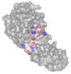
Localization of the active site residues on the surface of ColM. Representation in van der Waals surface of the D226KYDFNASTHR236 residues, localized at the surface of ColM. For these residues, a color code has been attributed to the atoms: carbon in white, nitrogen in blue and oxygen in red. The residues are identified through their position in the primary structure as well as through the corresponding one-letter code. The figure was prepared with the atomic coordinates from the PDB (PDB entry: 2XMX) by using PyMOL (DeLano Scientific).
Figure 8
Figure 8
3D-structures of Cmi. (a) Structure of Cmi as a monomer (PDB entry, 2XGL). The N-terminal (N-ter) and C-terminal (C-ter) ends are indicated. The cysteinyl residues 31 and 107 (Cys31 and Cys107), responsible for the formation of the disulfide bond, are colored in yellow and represented as sticks. (b) Structure of Cmi as a dimer (PDB entry, 4AEQ). Each protomer is colored differently (green for protomer A and blue for protomer B). The N-terminal (N-terA and N-terB) and C-terminal (C-terA and C-terB) of both protomers are indicated. The residues involved in the formation of intermolecular disulfide bridges are identified. Residues Cys31A and Cys107B, belonging to monomers A and B, respectively, form the first disulfide bridge. Residues Cys31B and Cys107A form the second one. The structures of Cmi as monomer and dimer are represented in cartoon. The figure was prepared with the atomic coordinates from the PDB by using PyMOL (DeLano Scientific).
Figure 9
Figure 9
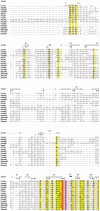
Sequence alignment of ColM and its homologs. The strictly conserved residues are highlighted in red, and the conserved residues in yellow. The strong sequence homology identified in the C-terminal part is boxed in red. The secondary structures of ColM are represented above the alignment. The amino acid residues essential for ColM activity are indicated in red below the sequences. This sequence alignment was generated with the ClustalW program and visualized thanks to the ESPript program [70].
Figure 9
Figure 9
Sequence alignment of ColM and its homologs. The strictly conserved residues are highlighted in red, and the conserved residues in yellow. The strong sequence homology identified in the C-terminal part is boxed in red. The secondary structures of ColM are represented above the alignment. The amino acid residues essential for ColM activity are indicated in red below the sequences. This sequence alignment was generated with the ClustalW program and visualized thanks to the ESPript program [70].
Figure 10
Figure 10
3D-structures of ColM-like proteins. PsyM (PDB entry: 4FZM), PaeM1 (PDB entry: 4G76), PcaM2 (stretched form; PDB entry: 4N58). For all structures, the translocation domain is colored in blue, the reception domain in green and the activity domain in red. 2Fe-2S: [2Fe-2S] cluster. The figure was prepared with the atomic coordinates from the PDB by using PyMOL (DeLano Scientific).

References

    1. Jack R.W., Tagg J.R., Ray B. Bacteriocins of gram-positive bacteria. Microbiol. Rev. 1995;59:171–200. doi: 10.1128/mr.59.2.171-200.1995. - DOI - PMC - PubMed
    1. Simons A., Alhanout K., Duval R.E. Bacteriocins, Antimicrobial Peptides from Bacterial Origin: Overview of Their Biology and Their Impact against Multidrug-Resistant Bacteria. Microorganisms. 2020;8:639. doi: 10.3390/microorganisms8050639. - DOI - PMC - PubMed
    1. Mavridou D., Gonzalez D., Kim W., West S.A., Foster K.R. Bacteria Use Collective Behavior to Generate Diverse Combat Strategies. Curr. Boil. 2018;28:345–355.e4. doi: 10.1016/j.cub.2017.12.030. - DOI - PubMed
    1. Granato E.T., Foster K.R. The Evolution of Mass Cell Suicide in Bacterial Warfare. Curr. Biol. 2020;30:2836–2843. doi: 10.1016/j.cub.2020.05.007. - DOI - PMC - PubMed
    1. Zacharof M.P., Lovitt R.W. Investigation of Shelf Life of Potency and Activity of the Lactobacilli Produced Bacteriocins Through Their Exposure to Various Physicochemical Stress Factors. Probiotics Antimicrob. Proteins. 2012;4:187–197. doi: 10.1007/s12602-012-9102-2. - DOI - PubMed

LinkOut - more resources