. 2021 Sep 21;10(9):1136.
doi: 10.3390/antibiotics10091136.

LyeTx I-b Peptide Attenuates Tumor Burden and Metastasis in a Mouse 4T1 Breast Cancer Model

Affiliations

LyeTx I-b Peptide Attenuates Tumor Burden and Metastasis in a Mouse 4T1 Breast Cancer Model

Mostafa A L Abdel-Salam et al. Antibiotics (Basel). .

Abstract

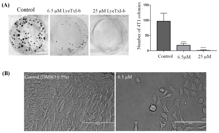
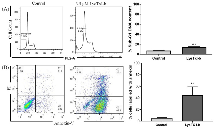
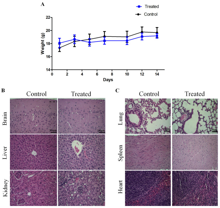
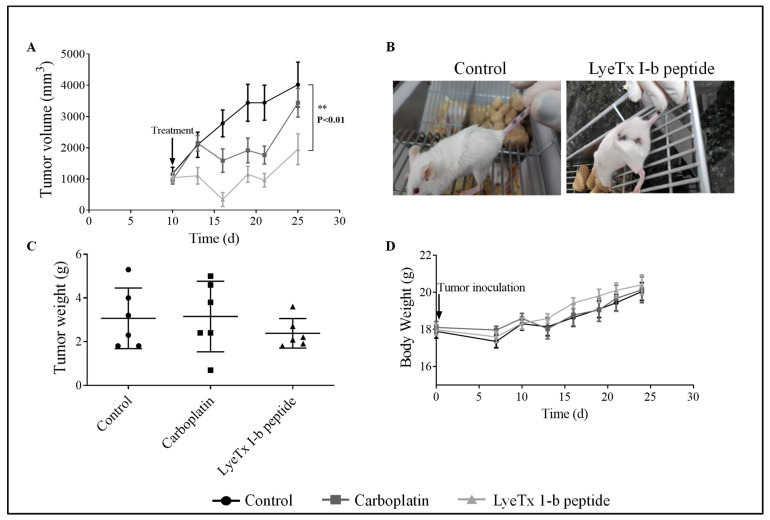
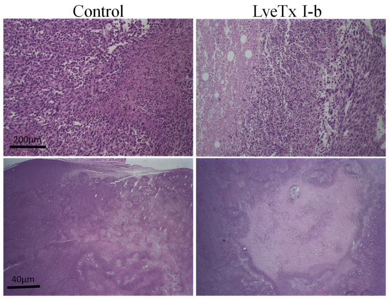
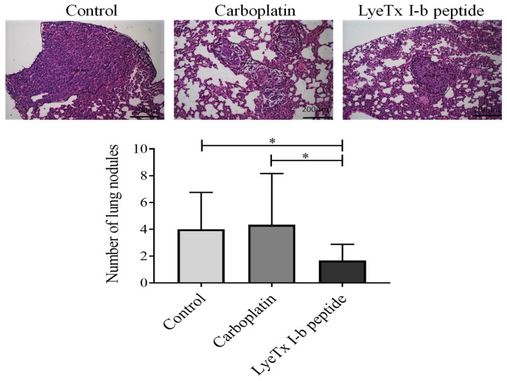
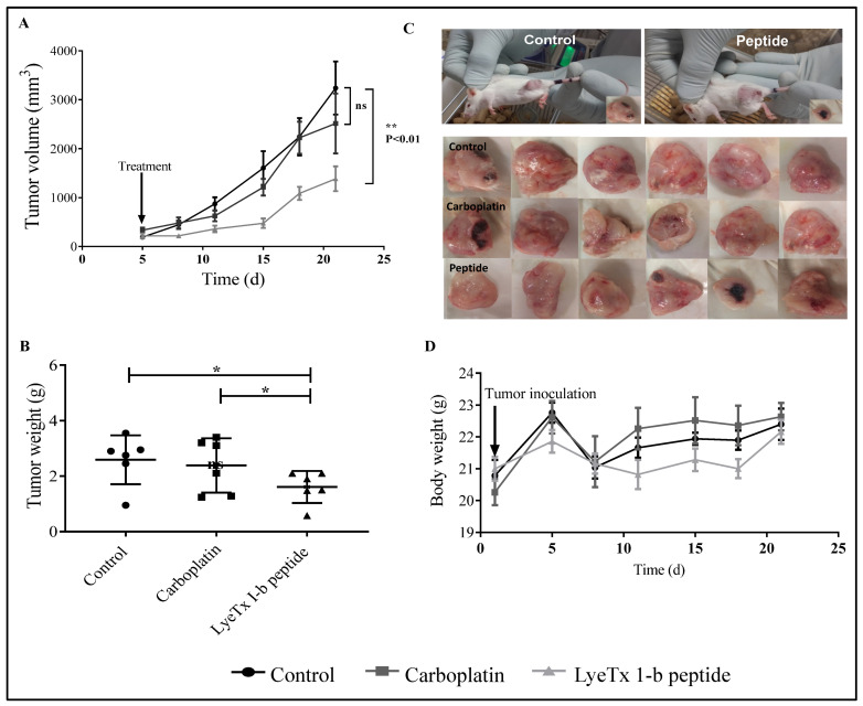
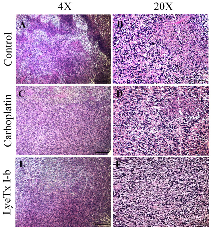
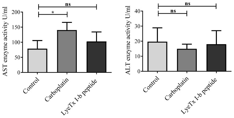
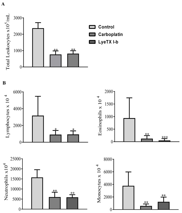
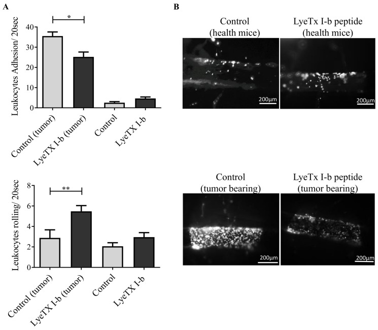
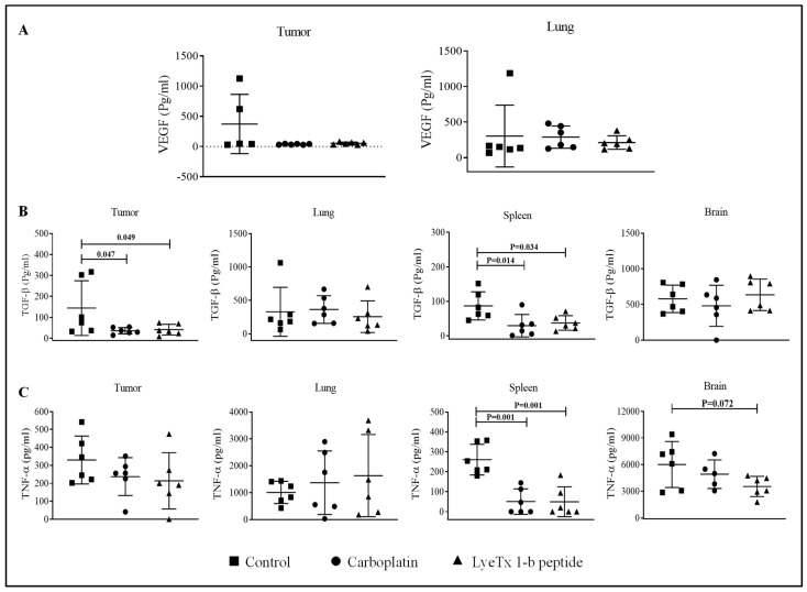
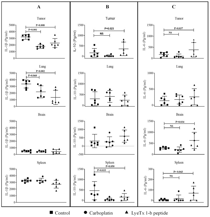
Cationic anticancer peptides have exhibited potent anti-proliferative and anti-inflammatory effects in neoplastic illness conditions. LyeTx I-b is a synthetic peptide derived from Lycosa erythrognatha spider venom that previously showed antibiotic activity in vitro and in vivo. This study focused on the effects of LyeTxI-b on a 4T1 mouse mammary carcinoma model. Mice with a palpable tumor in the left flank were subcutaneously or intratumorally injected with LyeTx I-b (5 mg/kg), which significantly decreased the tumor volume and metastatic nodules. Histological analyses showed a large necrotic area in treated primary tumors compared to the control. LyeTxI-b reduced tumor growth and lung metastasis in the 4T1 mouse mammary carcinoma model with no signs of toxicity in healthy or cancerous mice. The mechanism of action of LyeTx I-b on the 4T1 mouse mammary carcinoma model was evaluated in vitro and is associated with induction of apoptosis and cell proliferation inhibition. Furthermore, LyeTx I-b seems to be an efficient regulator of the 4T1 tumor microenvironment by modulating several cytokines, such as TGF-β, TNF-α, IL-1β, IL-6, and IL-10, in primary tumor and lung, spleen, and brain. LyeTx I-b also plays a role in leukocytes rolling and adhesion into spinal cord microcirculation and in the number of circulating leukocytes. These data suggest a potent antineoplastic efficacy ofLyeTx I-b.

Keywords: Lycosa erythrognatha; LyeTx I-b peptide; anticancer activity of LyeTx I-b; breast cancer; cytokines; immunomodulation; leukocyte recruitment.


Conflict of interest statement

The authors of this article certify that they have no conflict of interest.

Figures

Figure 1
Figure 1
Quantification of the survival of 4T1 single cell colonies after the treatment with LyeTx I-b (IC50 and IC80). The IC50 and IC80 were determined by the MTT method. Representative images of colonies of 4T1 after 10 days (A) and morphology of untreated (control) and treated cells with IC50 concentration of peptide (B) are shown. Data represent the mean ± SD of duplicates of three independent experiments, One-way ANOVA followed by Dunnett’s comparison test, **** p < 0.0001 compared to the control group.
Figure 2
Figure 2
(A) Representative histograms of cell cycle analysis after exposure to LyeTx I-b and carboplatin at IC50 values for 48 h. The results represent three independent experiments in triplicate and show significant differences between the negative control and the treated cells in terms of the G0/G1 (sub-diploid) population. (B) Illustrative histogram representing the quantification of the proportions of the dead and live cells after exposure of 4T1 cells to 6.5 µM of LyeTx I-b for 24 h and staining with annexin V/Propidium iodide (PI). The number of cells in each quadrant (Q) gate can be quantified as follows: Q4 = live cells (PI negative/annexin V negative); Q3 = apoptotic cells (PI negative/annexin V positive); Q2 = necrotic or late apoptotic cells (PI positive/annexin V positive); and, eventually, Q1 = nuclei without plasma membrane = (PI positive/annexin V negative). Three independent experiments in triplicate and unpaired t-test were performed, ** p < 0.01; *** p < 0.001 compared to the control group.
Figure 3
Figure 3
In vivo sub-acute toxicity after i.p. injection of LyeTxI-b peptide (5 mg/kg) (A) Effect of the peptide (5 mg/kg) on body weight of BALB/c mice over 14 days, n = 6. (B) Murine histopathological analyses of brain, liver, and kidney (C) Murine histopathological analyses of lung, spleen and heart. All tissues were stained with hematoxylin and eosin. Objective, 40×. Scale bar, 200 μm.
Figure 4
Figure 4
Effects of Tx I-b and carboplatin treatments (5 mg/kg) on 4T1 murine mammary carcinoma on the volume and weight of the tumor, and the body weight, n = 6. (A) LyeTx I-b decreased the tumor volume compared to the control group. (B) Representative images demonstrating a reduction in the size of tumor after treatment with LyeTx I-b. (C) LyeTx I-b reduced tumor weight. (D) No body weight differences over 25 days post-inoculation were observed on 4T1 murine mammary carcinoma model in the control, LyeTx I-b, and carboplatin groups. Data represent the mean ± SD, One-way ANOVA followed by Tukey’s multiple comparisons test, ** p < 0.01.
Figure 5
Figure 5
Assessment of morphological aspects of 4T1 murine primary tumor. Representative pictures of hematoxylin and eosin staining using low magnification showed a large necrosis area in the treated group compared to the untreated group, n = 6. In the control group, were observed inflammatory cell infiltrate, necrotic area, and viable tumor cells, whereas in the treated group the extensive necrotic area was characterized by the presence of nucleus chromatin condensation accompanied by fragmented nuclei into various segments (karyorrhexis). Objectives: 4× and 20×. Scale bars: 200 µm and 40 µm.
Figure 6
Figure 6
4T1 metastasis in the lung parenchyma. A significant reduction in the number of metastatic lesions in the LyeTx I-b group compared to the carboplatin treated and control groups was observed, n = 6. Hematoxylin and eosin (H&E) staining. Data represent the mean ± SD, One-way ANOVA followed by Tukey’s multiple comparisons test, * p < 0.05.
Figure 7
Figure 7
Effects of subcutaneous LyeTx I-b treatment on tumor volume, tumor weight, and body weight after 4T1 murine mammary carcinoma induction, n = 6. (A) LyeTx I-b (5 mg/kg) significantly decreased the tumor volume compared to the control and carboplatin (5 mg/kg). (B) LyeTx I-b reduced the tumor weight compared to the control and carboplatin groups. (C) Representative images demonstrating a reduction in the size of the tumor after the treatment with LyeTx I-b. (D) Influence of tumor induction and drug treatment on BALB/c body weight over 21 days post-inoculation of 4T1 tumor. Data represent the mean ± SD, One-way ANOVA followed by Tukey’s multiple comparisons test, * p < 0.05, ** p < 0.01. ns: means not statistically significant.
Figure 8
Figure 8
Histological effects of subcutaneous administration of LyeTx I-b on4T1 murine primary tumor, n = 6. (A,B) The control group exhibited the presence of neoplastic cells with a high degree of cellular polymorphism. (C,D) The carboplatin group showed inflammatory infiltrate characterized by mononuclear and neoplastic cells with a high degree of polymorphisms. (E,F) The reduction of areas of necrosis and fibrosis in the tumor periphery. (B,F) The group treated with LyeTx I-b presented a reduction in neoplastic cells and necrotic areas, associated with increased deposition of fibrous connective tissue. H&E staining. Objectives: 4× and 20×. Scale bars: 200 µm and 40 µm.
Figure 9
Figure 9
Plasma aspartate aminotransferase (AST) and alanine aminotransferase (ALT) activities in 4T1 tumor-bearing mice. Balb/c mice were injected subcutaneously 5 times, every 72 h, for 15 days with saline (control), LyeTX I-b (5 mg/kg), or carboplatin (5 mg/kg). AST and ALT levels were measured by using the colorimeter testing kit in triplicate, n = 6. Data represent the mean ± SD, One-way ANOVA followed by Tukey’s multiple comparisons test, * p < 0.05. ns: means not statistically significant.
Figure 10
Figure 10
Leukogram of peripheral blood counts in 4T1 tumor-bearing mice. The control received saline, and the other groups received carboplatin or LyeTx I-b (5 mg/kg), five doses every 72 h. (A) Total leukocytes count × 105. (B) Differential leukocytes count × 104, lymphocytes, eosinophils, neutrophils, and monocytes. LyeTx I-b and carboplatin induced leukopenia in treated groups. Data are presented as the mean ± SD, * p < 0.05; ** p < 0.01; *** p < 0.001 compared to the control group, n = 6 animals.
Figure 11
Figure 11
Evaluation of in vivo leukocyte recruitment by intravital microscopy, on day22 post-inoculation of 4T1 tumor cells. (A) Number of rolling and adhering leukocytes in health and tumor-bearing mice. Both control groups received 100 µL of saline, and treated groups received 5 mg/kg of LyeTx I-b dissolved in saline subcutaneously (5 doses/72 h apart). (B) Representative images of blood vessels of the spinal cord show the impact of LyeTx I-b treatment on leukocyte-endothelial interactions; higher numbers of adherent leukocytes in tumor-bearing control compared to the treated group were observed. Statistical analysis: Data are presented as the mean ± SD, * p < 0.05; ** p < 0.01, n = 6 animals. One-way ANOVA followed by Tukey’s multiple comparisons test.
Figure 12
Figure 12
Cytokines levels in the tumor LyeTx I-b attenuated VEGF, TGF-β, and TNF-α levels in the tumor microenvironment. Individual values of mice (pg/mL) are presented in a dot plot with an average of each group, n = 6. (A) Data correspond to VEGF differences found in primary tumor and lung. (B,C) TGF-β and TNF-α values in the primary tumor, brain, spleen, and lung. Statistical variation among groups was carried out in 6 animals of each group in duplicate using One-way ANOVA, p < 0.05.
Figure 13
Figure 13
LyeTx I-b changed levels of IL-1β, IL-6, and IL-10 expression in the tumor microenvironment. Individual values of mice (pg/mL) are presented in a dot plot with an average of each group, n = 6. (A) Data correspond to IL-1β found in the primary tumor, brain, spleen, and lung. (B,C) IL-10 and IL-6 levels, respectively. Statistical variation among groups was carried out in 6 animals of each group in duplicate using One-way ANOVA, p < 0.05. NS means not statistically significant.

Similar articles

Cited by

References

    1. Thomas M., Kelly E.D., Abraham J., Kruse M. Invasive lobular breast cancer: A review of pathogenesis, diagnosis, management, and future directions of early stage disease. Semin. Oncol. 2019;46:121–132. doi: 10.1053/j.seminoncol.2019.03.002. - DOI - PubMed
    1. Rahman M.M., Brane A.C., Tollefsbol T.O. MicroRNAs and Epigenetics Strategies to Reverse Breast Cancer. Cells. 2019;8:1214. doi: 10.3390/cells8101214. - DOI - PMC - PubMed
    1. World Health Organization . Breast Cancer. World Health Organization; Geneva, Switzerland: Mar 26, 2021.
    1. Sharma R. Breast cancer incidence, mortality and mortality-to-incidence ratio (MIR) are associated with human development, 1990–2016: Evidence from Global Burden of Disease Study 2016. Breast Cancer. 2019;26:428–445. doi: 10.1007/s12282-018-00941-4. - DOI - PubMed
    1. DeSantis C.E., Bray F., Ferlay J., Lortet-Tieulent J., Anderson B.O., Jemal A. International Variation in Female Breast Cancer Incidence and Mortality Rates. Cancer Epidemiol. Biomark. Prev. Publ. Am. Assoc. Cancer Res. Cosponsored Am. Soc. Prev. Oncol. 2015;24:1495–1506. doi: 10.1158/1055-9965.EPI-15-0535. - DOI - PubMed

LinkOut - more resources