Review
. 2021 Sep 10;13(18):4554.
doi: 10.3390/cancers13184554.

Oncologic Imaging of the Lymphatic System: Current Perspective with Multi-Modality Imaging and New Horizon

Affiliations
Review

Oncologic Imaging of the Lymphatic System: Current Perspective with Multi-Modality Imaging and New Horizon

Mohamed Elshikh et al. Cancers (Basel). .

Abstract

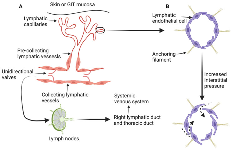
The lymphatic system is an anatomically complex vascular network that is responsible for interstitial fluid homeostasis, transport of large interstitial particles and cells, immunity, and lipid absorption in the gastrointestinal tract. This network of specially adapted vessels and lymphoid tissue provides a major pathway for metastatic spread. Many malignancies produce vascular endothelial factors that induce tumoral and peritumoral lymphangiogenesis, increasing the likelihood for lymphatic spread. Radiologic evaluation for disease staging is the cornerstone of oncologic patient treatment and management. Multiple imaging modalities are available to access both local and distant metastasis. In this manuscript, we review the anatomy, physiology, and imaging of the lymphatic system.

Keywords: conventional lymphography; lymphangiography; lymphatic metastasis; lymphatic system anatomy; lymphatic system imaging; lymphatic system physiology; lymphoscintigraphy; magnetic resonance lymphangiography.


Conflict of interest statement

The authors declare no conflict of interest.

Figures

Figure 1
Figure 1
Diagrammatic illustration of the anatomy and physiology of the lymphatic system. (A) The blind end lymphatic capillaries that drain in the pre-collecting lymphatic vessels. Lymph is then transferred to the collecting lymphatic vessels and get filtered by the lymph nodes. All lymph from the human body ends in the systemic venous system through either the thoracic duct or right lymphatic duct. (B) The effect on increased interstitial pressure on the loose junctions between lymphatic endothelial cells, leading to increased transmission of the interstitial fluid and proteins to the lymphatic capillaries and pre-collecting lymphatic vessels (dashed arrows). “Created by BioRender.com. accessed on 10 August 2021”.
Figure 2
Figure 2
Diagrammatic illustration of the superficial inguinal lymph nodes anatomy. The superficial inguinal lymph nodes are inferior to the inguinal ligament and divided in five groups by the greater saphenous vein (GSV) and a horizontal line through the saphenofemoral junction (SFJ). The five groups are superior medial (1), superior lateral (2), inferior lateral (3), inferior medial (4), and central group that overlies the SFJ. FV: femoral vein. The inferolateral group receives most of the lymphatic drainage of the lower extremity. “Created by BioRender.com. accessed on 10 August 2021”.
Figure 3
Figure 3
Axial contrast-enhanced computed tomography of the right hemipelvis demonstrates the right external iliac artery and veins with associated lateral (1), middle (2), and medial (3) external iliac lymph nodes. “Created by BioRander.com. accessed on 10 August 2021”.
Figure 4
Figure 4
Axial contrast-enhanced computed tomography of the pelvis shows the anterior internal iliac lymph node group (1) that follows the course of the internal iliac artery anterior division, the lateral sacral lymph node group (2) that runs along the lateral sacral artery, and the presacral lymph nodes (3) that lies in the midline presacral region. “Created by BioRander.com. accessed on 10 August 2021”.
Figure 5
Figure 5
Axial contrast-enhanced CT of the upper pelvis at the level of the common iliac vessels shows the common iliac artery and vein. The lateral common iliac lymph node subgroup (1) is located lateral to the common iliac artery, while the medial subgroup (2) is medial to the common iliac artery. The middle subgroup (3) is in the lumbosacral fossa. “Created by BioRender.com. accessed on 10 August 2021”.
Figure 6
Figure 6
Axial contrast-enhanced CT of the abdomen (A) demonstrates the distribution of the peri-aortic lymph nodes: 1; pre-aortic, 2; left lateral aortic, 3; right lateral aortic, and 4; post-aortic. Sagittal contrast-enhanced CT of the abdomen (B) shows the distribution of the pre-aortic lymph nodes along the origins of the visceral branches of the aorta (celiac, superior mesenteric, and the inferior mesenteric arteries). “Created by BioRender.com. accessed on 10 August 2021”.
Figure 7
Figure 7
Diagrammatic illustration of the lymphatic drainage of the whole human body. The right upper extremity, right hemiface, and right hemithorax are drained by the right lymphatic duct (green-colored). The thoracic duct drains the rest of the human body.
Figure 8
Figure 8
Ultrasound of a benign-appearing cervical lymph node (A) shows an oval hypoechoic lymph node with echogenic fatty hilum (arrow). Color doppler ultrasound of another benign-appearing lymph node (B) demonstrates a fatty hilum (normal) with mild hilar vascularity (arrow).
Figure 9
Figure 9
Ultrasound of the right axilla (A) demonstrates multiple, round, and hypoechoic metastatic lymph nodes with S/L axis ratio of approximately 1 and lack of the echogenic fatty hilum. Color Doppler ultrasound of the axillary lymph (B) nodes shows peripheral vascularity (arrow).
Figure 10
Figure 10
Doppler ultrasound of a right inguinal metastatic lymph node in a 67-year-old male with history of right ankle melanoma shows an enlarged hypoechoic lymph node with mixed (hilar and peripheral) vascularity.
Figure 11
Figure 11
A 61-year-old female patient with history of gastric cancer. PET/CT scan demonstrates diffuse thickening of the gastro-esophageal region as well as the gastric cardia (arrow) (A) consistent with gastric carcinoma, which is FDG-avid (arrow) with maximum SUV = 11.6 (B). There is no focal abnormal metabolic activity to suggest distant metastatic spread or regional lymphadenopathy.
Figure 12
Figure 12
A 77-year-old male patient with gastric cancer. PET/CT scan demonstrates marked thickening of the gastric antrum with increased uptake (A,B) consistent with gastric carcinoma, maximum SUV = 8.7 (B). Enlarging metastatic adenopathy in the gastrohepatic ligament is also noted (C,D), not hypermetabolic on PET/CT.
Figure 13
Figure 13
Axial fused PET-CT (A) and non-contrast CT of the abdomen shows mildly enlarged right paraaortic lymph node with increased FDG avidity on PET-CT (SUVmax 5.7), (B) biopsy proven Hodgkin lymphoma.
Figure 14
Figure 14
A 65-year-old male patient with right lower lobe lung cancer. The pulmonary mass seen in (A) shows avid FDG uptake (SUV = 4.6) in (C). Right hilar and subcarinal lymph nodes show mild FDG uptake (SUV = 2.6), which was found later to be inflammatory due to chemotherapy, resolving on (B,D). This is an example of false positive uptake in nonmalignant tissue.
Figure 15
Figure 15
A 75-year-old female patient with abdominal leiomyosarcoma and chylous ascites. (A) Conventional lymphangiography was performed through the right pedal approach. The right inguinal lymphatic vessels are opacified (arrow) with subsequent opacification of the right pelvic lymphatic network (B,C). Noncontrast CT performed after lymphangiography shows ascending opacification of the lymphatic vessels and cisterna chyle (D). Estimated site of leakage appears from small lymphatic vessels in the left peri-aortic region at level of L3 (E).
Figure 16
Figure 16
A 39-year-old male patient who underwent surgical resection of esophageal leiomyoma complicated by persistent chylous effusion. Successful percutaneous fluoroscopic-guided thoracic duct embolization using embolization coils. (A) Bilateral conventional lymphangiography was done through inguinal lymph nodes (arrowheads) with lymphatic vessels opacification (arrow). (B) Subsequent opacification of the lymphatic network and deep lymph nodes (arrow). (C) After 81 min, opacification of the cisterna chyle occurred (arrow). (D) Deployment of 3 embolization coils (white arrow) in the thoracic duct proximal to the estimated site of leak (arrowhead—surgical clips from previous operations).
Figure 17
Figure 17
A 68-year-old male with melanoma in the right temporal region who underwent lymphoscintigraphy (A) and complementary SPECT/CT scan in the coronal plane (B); (C,D) show CT (left) and fused SPECT/CT images (right) for sentinel lymph nodes mapping. Intradermal injection of the TC99m sulfur colloid in the right temple (arrow in (B)) shows tracer uptake in the pre-auricular region (arrowhead in (C)) and intra-parotid region (arrowhead in (D)), consistent with sentinel lymph drainage.
Figure 18
Figure 18
A 76-year-old male patient with a history of tongue base cancer status post-chemoradiation and left upper extremity lympho-venous bypass due to swelling of his left arm. Lymphoscintigraphy was performed by injection of sulfur colloid (radiotracer) into both hands. (A) The patient position. (B) Right anterior scintigraphy position with accumulation of tracer in both hands and tracer uptake in the right arm (black arrow). (C,D) Fused SPECT/CT with transit of tracer via proximal arm LN (white arrow in (C)) to reach lymph nodes at right axilla (white arrow (D)) with absence of any tracer uptake in the whole left upper extremity, confirming normal lymphatic drainage of the right arm with lymphedema of the left arm.

Similar articles

Cited by

References

    1. Al-Kofahi M., Yun J.W., Minagar A., Alexander J.S. Anatomy and roles of lymphatics in inflammatory diseases. Clin. Exp. Neuroimmunol. 2017;8:199–214. doi: 10.1111/cen3.12400. - DOI
    1. Scaglioni M.F., Suami H. Anatomy of the Lymphatic System and the Lymphosome Concept with Reference to Lymphedema. Semin. Plast. Surg. 2018;32:5–11. doi: 10.1055/s-0038-1635118. - DOI - PMC - PubMed
    1. Hsu M.C., Itkin M. Lymphatic Anatomy. Tech. Vasc. Interv. Radiol. 2016;19:247–254. doi: 10.1053/j.tvir.2016.10.003. - DOI - PubMed
    1. Tammela T., Alitalo K. Lymphangiogenesis: Molecular Mechanisms and Future Promise. Cell. 2010;140:460–476. doi: 10.1016/j.cell.2010.01.045. - DOI - PubMed
    1. Dellinger M.T., Witte M.H. Lymphangiogenesis, lymphatic systemomics, and cancer: Context, advances and unanswered questions. Clin. Exp. Metastasis. 2018;35:419–424. doi: 10.1007/s10585-018-9907-9. - DOI - PubMed

LinkOut - more resources