

PD-L1 Dependent Immunogenic Landscape in Hot Lung Adenocarcinomas Identified by Transcriptome Analysis
- PMID: 34572789
- PMCID: PMC8469831
- DOI: 10.3390/cancers13184562
PD-L1 Dependent Immunogenic Landscape in Hot Lung Adenocarcinomas Identified by Transcriptome Analysis
Abstract
Background: Lung cancer is the most frequent cause of cancer-related deaths worldwide. The clinical development of immune checkpoint blockade has dramatically changed the treatment paradigm for patients with lung cancer. Yet, an improved understanding of PD-1/PD-L1 checkpoint blockade-responsive biology is warranted.
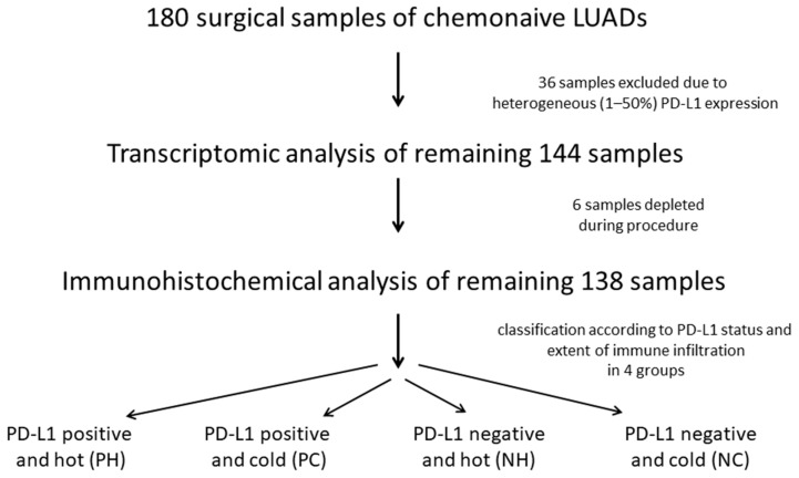
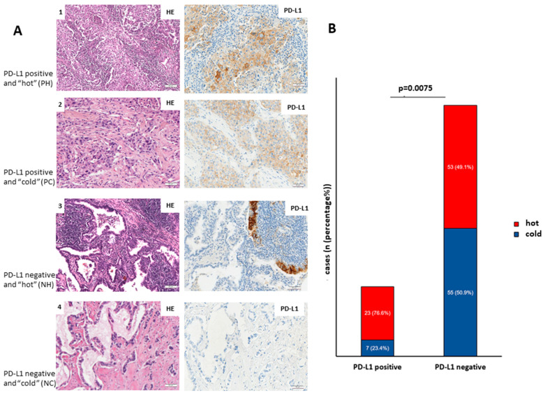
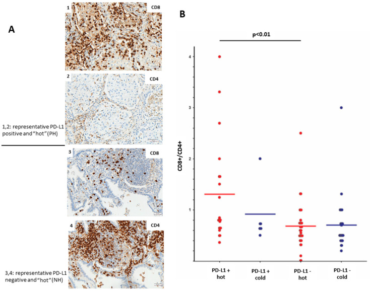
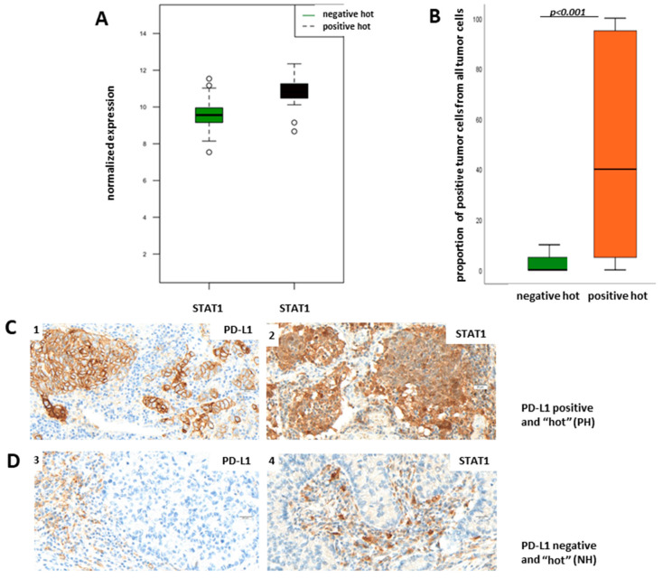
Methods: We aimed to identify the landscape of immune cell infiltration in primary lung adenocarcinoma (LUAD) in the context of tumoral PD-L1 expression and the extent of immune infiltration ("hot" vs. "cold" phenotype). The study comprises LUAD cases (n = 138) with "hot" (≥150 lymphocytes/HPF) and "cold" (<150 lymphocytes/HPF) tumor immune phenotype and positive (>50%) and negative (<1%) tumor PD-L1 expression, respectively. Tumor samples were immunohistochemically analyzed for expression of PD-L1, CD4, and CD8, and further investigated by transcriptome analysis.
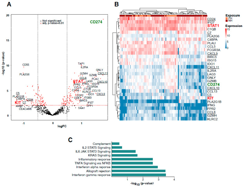
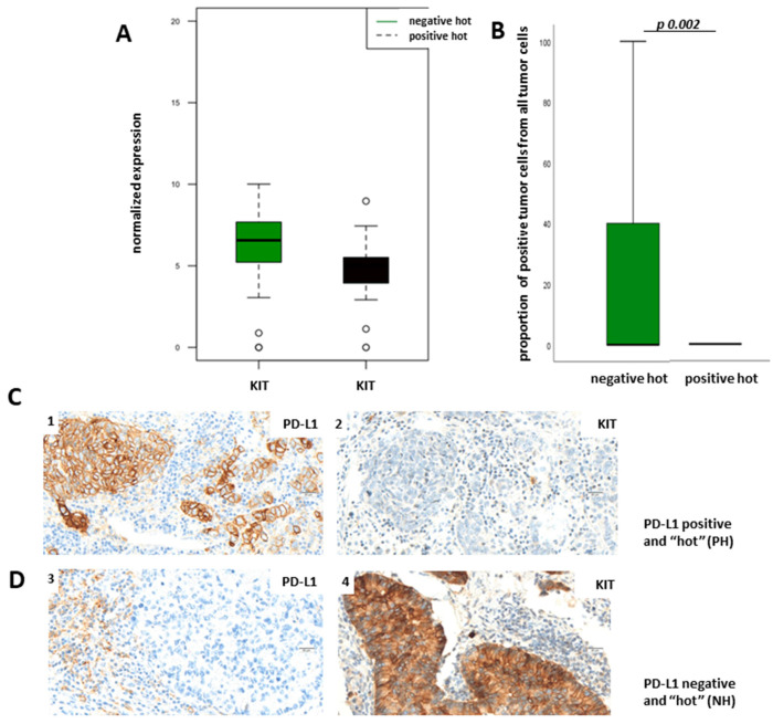
Results: Gene set enrichment analysis defined complement, IL-JAK-STAT signaling, KRAS signaling, inflammatory response, TNF-alpha signaling, interferon-gamma response, interferon-alpha response, and allograft rejection as significantly upregulated pathways in the PD-L1-positive hot subgroup. Additionally, we demonstrated that STAT1 is upregulated in the PD-L1-positive hot subgroup and KIT in the PD-L1-negative hot subgroup.
Conclusion: The presented study illustrates novel aspects of PD-L1 regulation, with potential biological relevance, as well as relevance for immunotherapy response stratification.
Keywords: cold; hot; immune phenotype; lung adenocarcinoma (LUAD); programmed cell death-ligand 1 (PD-L1); protein; transcriptome.
Conflict of interest statement
The authors declare no conflict of interest. The funders had no role in the design of the study; in the collection, analyses, or interpretation of data; in the writing of the manuscript, or in the decision to publish the results.
Figures
Similar articles
-
Gene landscape and correlation between B-cell infiltration and programmed death ligand 1 expression in lung adenocarcinoma patients from The Cancer Genome Atlas data set.PLoS One. 2018 Dec 6;13(12):e0208459. doi: 10.1371/journal.pone.0208459. eCollection 2018. PLoS One. 2018. PMID: 30521597 Free PMC article.
-
PD-L1 and PD-L2 expression correlated genes in non-small-cell lung cancer.Cancer Commun (Lond). 2019 Jun 3;39(1):30. doi: 10.1186/s40880-019-0376-6. Cancer Commun (Lond). 2019. PMID: 31159869 Free PMC article.
-
Programmed Death Ligand 1 Indicates Pre-Existing Adaptive Immune Response by Tumor-Infiltrating CD8+ T Cells in Non-Small Cell Lung Cancer.Int J Mol Sci. 2019 Oct 17;20(20):5138. doi: 10.3390/ijms20205138. Int J Mol Sci. 2019. PMID: 31627272 Free PMC article.
-
Potential biomarker for checkpoint blockade immunotherapy and treatment strategy.Tumour Biol. 2016 Apr;37(4):4251-61. doi: 10.1007/s13277-016-4812-9. Epub 2016 Jan 16. Tumour Biol. 2016. PMID: 26779629 Review.
-
The Next Immune-Checkpoint Inhibitors: PD-1/PD-L1 Blockade in Melanoma.Clin Ther. 2015 Apr 1;37(4):764-82. doi: 10.1016/j.clinthera.2015.02.018. Epub 2015 Mar 29. Clin Ther. 2015. PMID: 25823918 Free PMC article. Review.
Cited by
-
Cytochalasin H enhances sensitivity to gefitinib in non-small-cell lung cancer cells through inhibiting EGFR activation and PD-L1 expression.Sci Rep. 2024 Oct 25;14(1):25276. doi: 10.1038/s41598-024-76060-2. Sci Rep. 2024. PMID: 39455693 Free PMC article.
-
A novel diagnostic model for predicting immune microenvironment subclass based on costimulatory molecules in lung squamous carcinoma.Front Genet. 2022 Dec 14;13:1078790. doi: 10.3389/fgene.2022.1078790. eCollection 2022. Front Genet. 2022. PMID: 36588791 Free PMC article.
-
Deciphering MOSPD1's impact on breast cancer progression and therapeutic response.Biol Direct. 2024 Oct 5;19(1):88. doi: 10.1186/s13062-024-00531-9. Biol Direct. 2024. PMID: 39369222 Free PMC article.
References
-
- Freeman G.J., Long A.J., Iwai Y., Bourque K., Chernova T., Nishimura H., Fitz L.J., Malenkovich N., Okazaki T., Byrne M.C., et al. Engagement of the PD-1 Immunoinhibitory Receptor by a Novel B7 Family Member Leads to Negative Regulation of Lymphocyte Activation. J. Exp. Med. 2000;192:1027–1034. doi: 10.1084/jem.192.7.1027. - DOI - PMC - PubMed
Grants and funding
LinkOut - more resources
Full Text Sources
Molecular Biology Databases
Research Materials
Miscellaneous
