Review
. 2021 Sep 12;13(18):4578.
doi: 10.3390/cancers13184578.

EBV-Driven Lymphoproliferative Disorders and Lymphomas of the Gastrointestinal Tract: A Spectrum of Entities with a Common Denominator (Part 1)

Affiliations
Review

EBV-Driven Lymphoproliferative Disorders and Lymphomas of the Gastrointestinal Tract: A Spectrum of Entities with a Common Denominator (Part 1)

Magda Zanelli et al. Cancers (Basel). .

Abstract

EBV is the most common persistent virus in humans. The interaction of EBV with B lymphocytes, which are considered the virus reservoir, is at the base of the life-long latent infection. Under circumstances of immunosuppression, the balance between virus and host immune system is altered and hence, EBV-associated lymphoid proliferations may originate. These disorders encompass several entities, ranging from self-limited diseases with indolent behavior to aggressive lymphomas. The virus may infect not only B-cells, but even T- and NK-cells. The occurrence of different types of lymphoid disorders depends on both the type of infected cells and the state of host immunity. EBV-driven lymphoproliferative lesions can rarely occur in the gastrointestinal tract and may be missed even by expert pathologists due to both the uncommon site of presentation and the frequent overlapping morphology and immunophenotypic features shared by different entities. The aim of this review is to provide a comprehensive overview of the current knowledge of EBV-associated lymphoproliferative disorders, arising within the gastrointestinal tract. The review is divided in three parts. In this part, the available data on EBV biology, EBV-positive mucocutaneous ulcer, EBV-positive diffuse large B-cell lymphoma, not otherwise specified and classic Hodgkin lymphoma are discussed.

Keywords: EBV-positive; EBV-positive mucocutaneous ulcer; Epstein–Barr virus; classic Hodgkin lymphoma; diffuse large B-cell lymphoma; not otherwise specified.


Conflict of interest statement

The authors declare no conflict of interest.

Figures

Figure 1
Figure 1
(A) EBV-positive DLBCL, NOS: medium power view showing a polymorphic infiltrate within the gastric mucosa (Hematoxylin and eosin, magnification 400×; original image from Prof S.A. (B) EBV-positive DLBCL, NOS: CD79alpha positivity highlighting the B-cell phenotype of the lymphoid infiltrate (immunostaining, magnification 200×; original image from Prof S.A.).
Figure 2
Figure 2
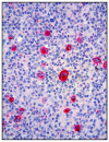
(A) EBV-positive DLBCL, NOS: Ki67 immunostaining revealing an elevated proliferative index of the lymphoid infiltrate (magnification 200×; original image from Prof S.A.); (B) EBV-positive DLBCL, NOS: EBER showing positive signals for EBV in the nuclei of the lymphoid infiltrate (inside the red circle) (magnification 200×; original image from Prof S.A.).
Figure 3
Figure 3
(A) cHL: medium power view showing a dense lymphoid infiltrate involving the small bowel wall (Hematoxylin and eosin, magnification 100×, original image from Prof S.A.); (B) cHL: high power of the polymorphic lymphoid infiltrate containing large-sized cells with Hodgkin features (Hematoxylin and eosin, magnification 200×; original image from Prof S.A.).
Figure 4
Figure 4
(A) cHL: CD15 expression highlighting a Hodgkin cell with membranous positivity (inside the blue circle); in the background, granulocytes are strongly CD15 positive (immunostaining, magnification 400×; original image from Prof S.A.); (B) cHL: weak PAX5 positivity of a Hodgkin cell, inside the blue circle; unlike Hodgkin cells, B lymphocytes strongly express PAX5 (immunostaining, magnification 400×; original image from Prof S.A.).
Figure 5
Figure 5
cHL: LMP1 expression in Hodgkin cells (immunostaining, magnification 400×; original image from Prof S.A.).

References

    1. Taylor G.S., Long H.M., Brooks J.M., Rickinson A.B., Hislop A.D. The immunology of Epstein-Barr virus induced disease. Annu. Rev. Immunol. 2015;33:787–821. doi: 10.1146/annurev-immunol-032414-112326. - DOI - PubMed
    1. Young L.S., Yap L.F., Murray P.G. Epstein-Barr virus: More than 50 years old and still providing surprises. Nat. Rev. Cancer. 2016;16:789–802. doi: 10.1038/nrc.2016.92. - DOI - PubMed
    1. Resk S.A., Weiss L.M. Epstein-Barr virus-associated lymphoproliferative disorders. Hum. Pathol. 2007;38:1293–1304. doi: 10.1016/j.humpath.2007.05.020. - DOI - PubMed
    1. Tanner J.E., Alfieri C. The Epstein-Barr virus and post-transplant lymphoproliferative disease: Interplay of immunosuppression, EBV, and the immune system in disease pathogenesis. Transpl. Infect. Dis. 2001;3:60–69. doi: 10.1034/j.1399-3062.2001.003002060.x. - DOI - PubMed
    1. Hakim F.T., Gress R.E. Immunosenescence: Deficits in adaptative immunity in the elderly. Tissue Antigens. 2007;70:179–189. doi: 10.1111/j.1399-0039.2007.00891.x. - DOI - PubMed

Grants and funding

LinkOut - more resources