. 2021 Aug 26;10(9):1363.
doi: 10.3390/antiox10091363.

p47phox-Dependent Oxidant Signalling through ASK1, MKK3/6 and MAPKs in Angiotensin II-Induced Cardiac Hypertrophy and Apoptosis

Affiliations

p47phox-Dependent Oxidant Signalling through ASK1, MKK3/6 and MAPKs in Angiotensin II-Induced Cardiac Hypertrophy and Apoptosis

Fangfei Liu et al. Antioxidants (Basel). .

Abstract

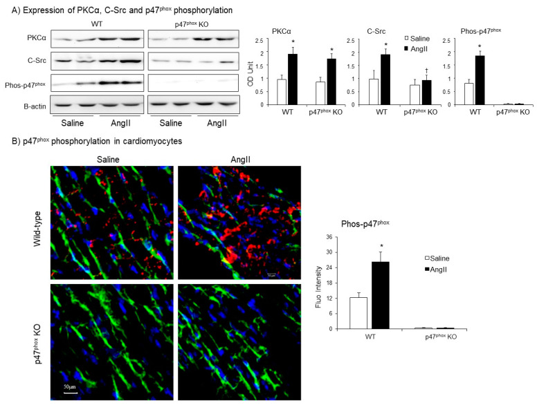
The p47phox is a key regulatory subunit of Nox2-containing NADPH oxidase (Nox2) that by generating reactive oxygen species (ROS) plays an important role in Angiotensin II (AngII)-induced cardiac hypertrophy and heart failure. However, the signalling pathways of p47phox in the heart remains unclear. In this study, we used wild-type (WT) and p47phox knockout (KO) mice (C57BL/6, male, 7-month-old, n = 9) to investigate p47phox-dependent oxidant-signalling in AngII infusion (0.8 mg/kg/day, 14 days)-induced cardiac hypertrophy and cardiomyocyte apoptosis. AngII infusion resulted in remarkable high blood pressure and cardiac hypertrophy in WT mice. However, these AngII-induced pathological changes were significantly reduced in p47phox KO mice. In WT hearts, AngII infusion increased significantly the levels of superoxide production, the expressions of Nox subunits, the expression of PKCα and C-Src and the activation of ASK1 (apoptosis signal-regulating kinase 1), MKK3/6, ERK1/2, p38 MAPK and JNK signalling pathways together with an elevated expression of apoptotic markers, i.e., γH2AX and p53 in the cardiomyocytes. However, in the absence of p47phox, although PKCα expression was increased in the hearts after AngII infusion, there was no significant activation of ASK1, MKK3/6 and MAPKs signalling pathways and no increase in apoptosis biomarker expression in cardiomyocytes. In conclusion, p47phox-dependent redox-signalling through ASK1, MKK3/6 and MAPKs plays a crucial role in AngII-induced cardiac hypertrophy and cardiomyocyte apoptosis.

Keywords: Angiotensin II; apoptosis; cardiac hypertrophy; knockout mice; p47phox; redox-signalling.


Conflict of interest statement

The authors declare no conflict of interest.

Figures

Figure 1
Figure 1
Development of hypertension and cardiac hypertrophy in AngII-infused WT and p47phox KO mice. (A) Systolic blood pressure. (B) Diastolic blood pressure. Day 0: day of minipump implantation. Day 14: day of minipump removal. (C) Heart weights. (D) Heart weight (HW, mg)/body weight (BW, g) ratio. (E) Left panels: Representative images of cardiomyocyte sizes on the cross-sections of left ventricular tissues. The cardiomyocytes membranes were labelled with WGA-FITC (green). Right panel: Statistical analysis of cardiomyocyte cross-sectional areas (µm2). n = 9 mice per group. Statistical analyses were performed using two-way ANOVA. * p < 0.05 for AngII values versus saline values in the same genetic group;  p < 0.05, for p47phox KO AngII values versus WT AngII values.
Figure 2
Figure 2
Cardiac ROS production. (A) Levels of O2●− production measured by lucigenin-chemiluminescence. Left panel: Representative examples of kinetic measurements of O2●− production by WT heart homogenates. NADPH (0.1 mM) was added at 10 min. Tiron (10 mM) was added at 30 min to scavenge O2●−. Right panel: Differences in NADPH-dependent O2●− production measured between 10–30 min shown in the left panel. (B) The effects of different enzyme inhibitors on the levels of O2●− by AngII-infused WT heart homogenates. L-NAME: (NG-nitroarginine methyl ester), NOS inhibitor; Rotenone: mitochondrial respiratory chain inhibitor; Oxypurinol, xanthine oxidase inhibitor; DPI: (diphenyleneiodonium), flavoprotein inhibitor; SOD: (superoxide dismutase). (C) Cardiac H2O2 production detected by amplex red assay. (D) In situ detection of reactive oxygen species production by DHE fluorescence. Left panel: Representative images of DHE staining on cardiac sections; right panel: Quantification of DHE fluorescence intensity. n = 9 per group. Statistical comparisons were done using one-way ANOVA for inhibitor assay, and two-way ANOVA for the rests (panels A, C, D). * p < 0.05 for indicated AngII values versus saline values in the same genetic group; p < 0.05 for p47phox KO AngII values versus WT AngII values.
Figure 3
Figure 3
Expressions of isoforms of the catalytic subunit of Nox (i.e., Nox1. Nox2, Nox4) and other subunits of Nox (p47phox, p22phox, p67phox and rac1) in murine hearts. Left panels: Representative Western blot images. β-actin detected in the same samples were used as loading controls. Right panels: Optical densities (OD) of Western blot bands were quantified and normalised to the levels of β-actin detected in the same samples. n = 9 per group. Statistical comparisons were made using two-way ANOVA. * p < 0.05 for AngII values versus saline values in the same genetic group; p < 0.05 for p47phox KO AngII values versus WT AngII values.
Figure 4
Figure 4
Expressions of protein kinase C alpha (PKCα), Proto-oncogene tyrosine-protein kinase Src (C-Src) and p47phox phosphorylation in AngII-infused murine hearts. (A) Western blots. Left panels: Representative images. β-actin detected in the same samples were used as loading controls. Right panels: Optical densities (OD) of Western blot bands were quantified and normalised to the levels of β-actin detected in the same samples. (B) Confocal immunofluorescence of cardiac sections. Left panel: Representative immunofluorescence images. Cardiomyocyte cell membrane was labelled by WGA-FITC (green) and p47phox phosphorylation was identified using phos-p47phox specific antibody (Cy3, red). Nuclei were labelled by DAPI (blue). Right panel: Quantification of phos-p47phox fluorescence intensity. n = 9 hearts per group. Data were presented as Mean ± SD. Statistical comparisons were made using two-way ANOVA. * p < 0.05 for AngII values versus saline values in the same genetic group.
Figure 5
Figure 5
AngII-induced activation of mitogen-activated protein kinase kinase 3/6 (MKK3/6), mitogen-activated protein kinases (i.e., ERK1/2, p38MAPK and JNK) and Akt (also called protein kinase B) in murine hearts. Left panels: Representative immunoblotting images. The total protein bands of each molecule in heart homogenates were pre-tested for equal loading. Right panels: Quantification of the optical densities (OD) of phos-protein bands expressed as phosphorylated/total (P/T) protein ratio. n = 9 mice per group. Data were presented as Mean ± SD. Statistical comparisons were made using two-way ANOVA. * p < 0.05 for AngII values versus saline values in the same genetic group.  p < 0.05 for p47phox KO AngII values versus WT AngII values.
Figure 6
Figure 6
Activation of apoptosis signal-regulating kinase 1 (ASK1), p53 and phosphorylation of H2A histone family member X (γH2AX) in murine hearts. (A) Western blots for the expressions of phos-ASK1, p53 and γH2AX. Optical densities of protein bands were quantified and normalised to the levels of β-actin detected in the same samples. (B) Confocal immunofluorescence detection of Nox2 (Cy3 labelled, red) and phos-ASK1 expressions (FITC labelled, green) in the cardiac sections. (C) Confocal immunofluorescence detection of γH2AX expression (Cy3-labelled, red) in the cardiac sections. The cardiomyocyte membrane was labelled by WGA-FITC (green), and the nuclei were labelled by DAPI (blue). The specific fluorescent densities were quantified. n = 9 mice per group. Data were presented as Mean ± SD. Statistical comparisons were made using two-way ANOVA. * p < 0.05, for AngII values versus saline values in the same genetic group. p < 0.05 for p47phox KO AngII values versus WT AngII values.
Figure 7
Figure 7
Schematic illustration of p47phox redox-signalling pathways examined.

References

    1. Lassegue B., Martin A.S., Griendling K.K. Biochemistry, physiology, and pathophysiology of NADPH oxidases in the cardiovascular system. Circ. Res. 2012;110:1364–1390. doi: 10.1161/CIRCRESAHA.111.243972. - DOI - PMC - PubMed
    1. Forrester S.J., Booz G.W., Sigmund C.D., Coffman T.M., Kawai T., Rizzo V., Scalia R., Eguchi S. Angiotensin II Signal Transduction: An Update on Mechanisms of Physiology and Pathophysiology. Physiol. Rev. 2018;98:1627–1738. doi: 10.1152/physrev.00038.2017. - DOI - PMC - PubMed
    1. Fan L.M., Douglas G., Bendall J.K., McNeill E., Crabtree M.J., Hale A.B., Mai A., Li J.M., McAteer M.A., Schneider J.E., et al. Endothelial cell-specific reactive oxygen species production increases susceptibility to aortic dissection. Circulation. 2014;129:2661–2672. doi: 10.1161/CIRCULATIONAHA.113.005062. - DOI - PMC - PubMed
    1. Fan L.M., Geng L., Cahill-Smith S., Liu F., Douglas G., McKenzie C.A., Smith C., Brooks G., Channon K.M., Li J.M. Nox2 contributes to age-related oxidative damage to neurons and the cerebral vasculature. J. Clin. Investig. 2019;129:3374–3386. doi: 10.1172/JCI125173. - DOI - PMC - PubMed
    1. Borchi E., Bargelli V., Stillitano F., Giordano C., Sebastiani M., Nassi P.A., d’Amati G., Cerbai E., Nediani C. Enhanced ROS production by NADPH oxidase is correlated to changes in antioxidant enzyme activity in human heart failure. Biochim. Biophys. Acta. 2010;1802:331–338. doi: 10.1016/j.bbadis.2009.10.014. - DOI - PubMed

LinkOut - more resources