. 2021 Aug 28;10(9):1373.
doi: 10.3390/antiox10091373.

Antioxidant, Anti-Inflammatory, and Anti-Aging Potential of a Kalmia angustifolia Extract and Identification of Some Major Compounds

Affiliations

Antioxidant, Anti-Inflammatory, and Anti-Aging Potential of a Kalmia angustifolia Extract and Identification of Some Major Compounds

Alexe Grenier et al. Antioxidants (Basel). .

Abstract

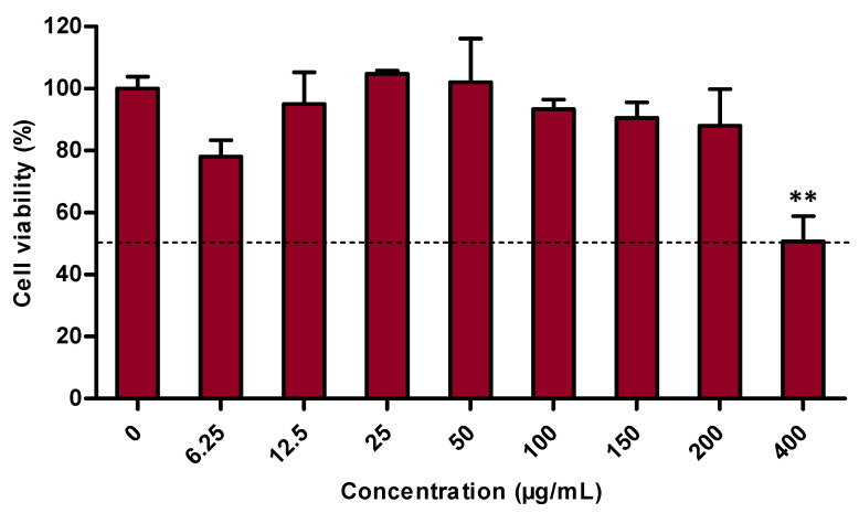
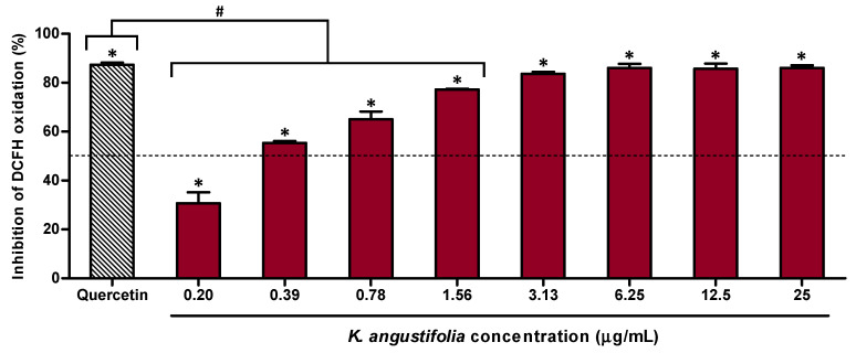
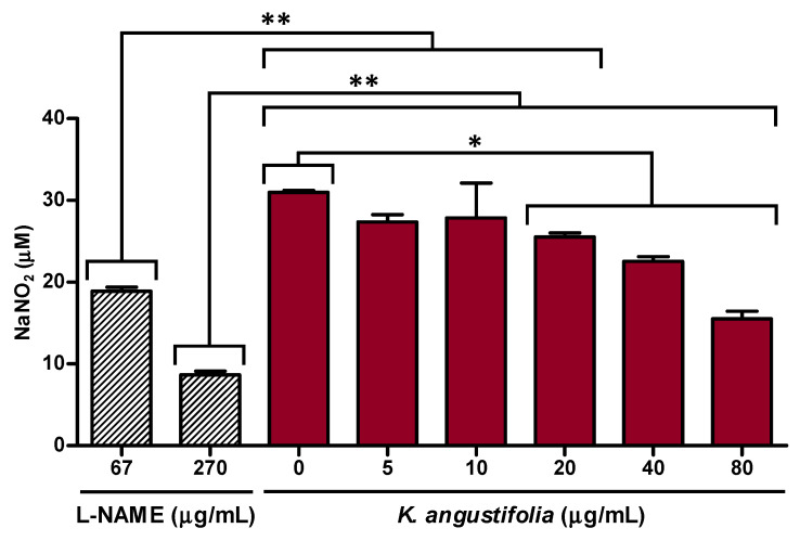
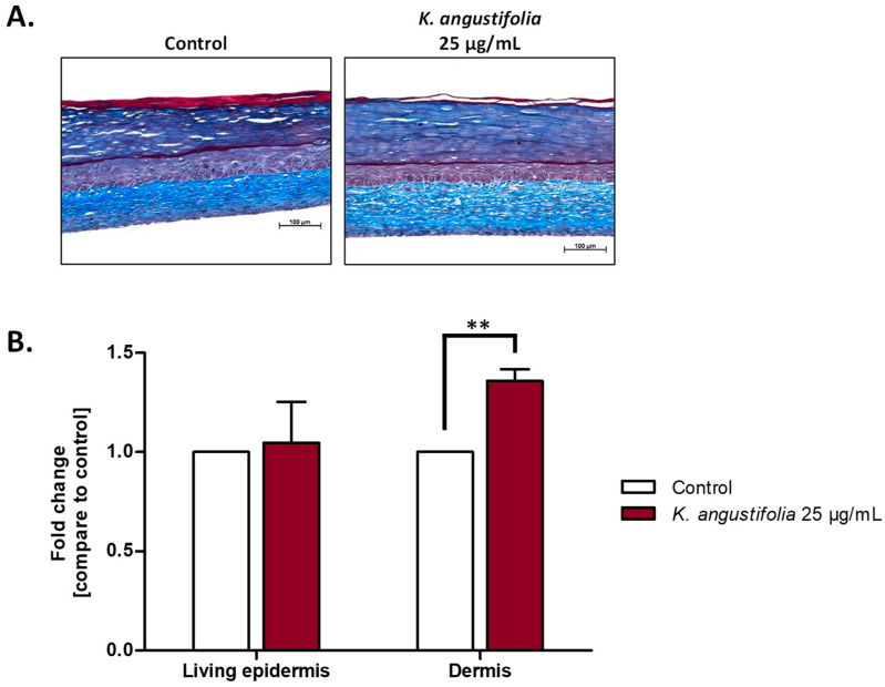
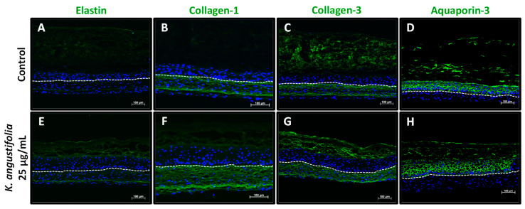
Skin aging is the most visible element of the aging process, giving rise to a major concern for many people. Plants from the Ericaceae family generally have antioxidant and anti-inflammatory properties, making them potential anti-aging active ingredients. This study aimed to evaluate the safety and anti-aging efficacy of a Kalmia angustifolia extract using reconstructed skin substitutes. The safety evaluation was performed using a 3-(4,5-dimethylthiazolyl-2)-2,5-diphenyltetrazolium bromide (MTT) assay, while the efficacy was determined by assessing antioxidant and anti-inflammatory activity and analyzing skin substitutes reconstructed according to the self-assembly method by histology and immunofluorescence staining (elastin, collagen-1, collagen-3, aquaporin-3). The cell viability assay established the safety of the extract at a concentration up to 200 μg/mL. The Oxygen Radical Absorbance Capacity (ORAC) assay and a cell-based assay using 2',7'-dichlorofluorescein-diacetate (DCFH-DA) revealed a strong antioxidant activity with an ORAC value of 16 µmol Trolox Equivalent/mg and a half-maximal inhibitory concentration (IC50) of 0.37 ± 0.02 μg/mL, while an interesting anti-inflammatory activity was found in the inhibition of NO production, with an inhibition percentage of NO production of 49 ± 2% at 80 µg/mL. The isolation and characterization of the extract allowed the identification of compounds that could be responsible for these biological activities, with two of them being identified for the first time in K. angustifolia: avicularin and epicatechin-(2β-O-7, 4β-6)-ent-epicatechin. Histological analyses of skin substitutes treated with the extract showed an increase in dermal thickness compared with the controls. K. angustifolia extract enhanced the expression of elastin and collagen-1, which are usually decreased with skin aging. These results suggest that K. angustifolia has promising antioxidant efficacy and anti-aging potential.

Keywords: Sheep Laurel; active ingredients; anti-inflammatory activity; antioxidant activity; natural products; skin aging; skin substitutes; tissue engineering.


Conflict of interest statement

The authors declare no conflict of interest.

Figures

Figure 1
Figure 1
Cytotoxicity assessment of the K. angustifolia extract on primary human keratinocytes performed by measuring metabolic activity with a MTT assay. All the experiments were carried out in at least triplicate and presented results are representative of at least two different experiments (N = 2, n = 3). Data are presented as means of the triplicates ± S.D. The horizontal line is set at 50% cell viability. Statistical significance was determined using a one-way ANOVA followed by a Dunnett’s post hoc test (p-value < 0.05 compared to control), ** p-value < 0.01.
Figure 2
Figure 2
Antioxidant activity of the K. angustifolia extract on the human skin fibroblast WS1 cell line treated with tert-butyl hydroperoxide (t-BuOOH), determined by a cell-based assay using DCFH-DA. All the experiments were carried out in at least triplicate and presented results are representative of at least two different experiments (N = 2, n = 3). Data are presented as means of the triplicates ± S.D. The horizontal line is set at 50% inhibition. Statistical significance was determined using a one-way ANOVA followed by a Dunnett’s post hoc test (p-value < 0.05 compared to control of cells treated with only t-BuOOH (200 µM); * p-value < 0.05) or Bonferroni’s post hoc test (p-value < 0.05 compared to positive control quercetin (1.56 µg/mL); # p-value < 0.001).
Figure 3
Figure 3
K. angustifolia extract inhibits NO overproduction in LPS-stimulated RAW 264.7 macrophages. All the experiments were carried out in triplicate and presented results are representative of at least two different experiments (N = 2, n = 3). Data are presented as means of the triplicates ± S.D. Statistical significance was determined using a one-way ANOVA followed by a Bonferroni’s post hoc test (p-value < 0.05 compared to controls (L-NAME: 67 and 270 µg/mL, or K. angustifolia: 0 µg/mL)), * p-value < 0.05, ** p-value < 0.01. L-NAME (N-ω-nitro-arginine methyl ester hydrochloride), at 67 μg/mL (250 μM) and 270 μg/mL (1 mM), was used as positive control.
Figure 4
Figure 4
Effect of the K. angustifolia extract on reconstructed skin substitutes histology. (A) Histological analyses of Masson’s trichrome stained skin substitute sections. Stratum corneum in dark blue, living epidermis in violet and dermis in light blue. Objective 10×, scale bar: 100 μm. (B) Fold change in the thickness of the living epidermis and dermis. Fold change is defined as the ratio of treated substitutes’ thickness value to the control (without treatment) thickness value. Two substitutes for each condition were analyzed and confirmed with three different cell populations (N = 3, n = 6). Data are presented as means of the three different cell populations ± S.D. Statistical significance was determined using a t-test, ** p-value < 0.01.
Figure 5
Figure 5
Aging markers visualized by immunofluorescence staining. Elastin (A,E), collagen-1 (B,F), collagen-3 (C,G) and aquaporin-3 (D,H) expression, which usually decreases during skin aging, is shown in healthy skin substitutes untreated (AD) and treated with the K. angustifolia extract at 25 μg/mL (EH). The nuclei were stained with DAPI (blue). The dotted line represents the separation between the epidermis and dermis. Two substitutes for each condition were analyzed and confirmed with three different cell populations (N = 3, n = 2; scale bar: 100 μm).

References

    1. Zhang S., Duan E. Fighting against Skin Aging: The Way from Bench to Bedside. Cell Transpl. 2018;27:729–738. doi: 10.1177/0963689717725755. - DOI - PMC - PubMed
    1. Tobin D.J., Veysey E.C., Finlay A.Y. Aging and the Skin. In: Fillit H.M., Rockwood K., Young J.B., editors. Textbook of Geriatric Medicine and Gerontology. Elsevier; London, UK: 2016.
    1. Titus B.D., Sidhu S.S., Mallik A.U. A summary of Some Studies on Kalmia angustifolia L.: A Problem Species in Newfoundland Forestry. Government of Canada, Natural Resources Canada, Canadian Forest Service; St. John’s, NL, Canada: 1995. pp. 1–68.
    1. Erichsen-Brown C. Medicinal and Other Uses of North American Plants: A Historical Survey with Special Reference to the Eastern Indian Tribes. Dover Publications; Mineola, NY, USA: 1989. p. 544.
    1. Jansen S.A., Kleerekooper I., Hofman Z.L.M., Kappen I.F.P.M., Stary-Weinzinger A., van der Heyden M.A.G. Grayanotoxin poisoning: ‘mad honey disease’ and beyond. Cardiovasc. Toxicol. 2012;12:208–215. doi: 10.1007/s12012-012-9162-2. - DOI - PMC - PubMed