Review
. 2021 Sep 9;10(9):1440.
doi: 10.3390/antiox10091440.

Resveratrol as an Adjunctive Therapy for Excessive Oxidative Stress in Aging COVID-19 Patients

Affiliations
Review

Resveratrol as an Adjunctive Therapy for Excessive Oxidative Stress in Aging COVID-19 Patients

Min-Tser Liao et al. Antioxidants (Basel). .

Abstract

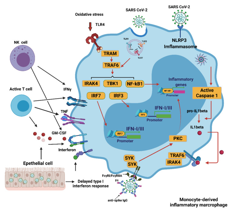
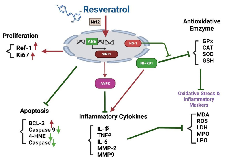
The coronavirus disease 2019 (COVID-19) pandemic continues to burden healthcare systems worldwide. COVID-19 symptoms are highly heterogeneous, and the patient may be asymptomatic or may present with mild to severe or fatal symptoms. Factors, such as age, sex, and comorbidities, are key determinants of illness severity and progression. Aging is accompanied by multiple deficiencies in interferon production by dendritic cells or macrophages in response to viral infections, resulting in dysregulation of inflammatory immune responses and excess oxidative stress. Age-related dysregulation of immune function may cause a more obvious pathophysiological response to SARS-CoV-2 infection in elderly patients and may accelerate the risk of biological aging, even after recovery. For more favorable treatment outcomes, inhibiting viral replication and dampening inflammatory and oxidative responses before induction of an overt cytokine storm is crucial. Resveratrol is a potent antioxidant with antiviral activity. Herein, we describe the reasons for impaired interferon production, owing to aging, and the impact of aging on innate and adaptive immune responses to infection, which leads to inflammation distress and immunosuppression, thereby causing fulminant disease. Additionally, the molecular mechanism by which resveratrol could reverse a state of excessive basal inflammatory and oxidative stress and low antiviral immunity is discussed.

Keywords: SARS-CoV-2; adaptive immunity; aging; antioxidants; inflammation; innate immunity; oxidative stress; resveratrol.


Conflict of interest statement

The authors declare no conflict of interest.

Figures

Figure 1
Figure 1
Activation of monocyte-derived macrophages in COVID-19. Several mechanisms induce the excessive activation of monocytes/macrophages during a SARS-CoV-2 infection. The delayed production of IFN I results in the continued conscription of circulating monocytes into the pulmonary parenchyma. Activated NK and T cells also favor infiltrating cells derived from monocytes. Virus detection may trigger the activation of TLR7 as a result of the recognition of the viral single-stranded RNA. IFN I increases the expression of the SARS-CoV-2 entry receptor ACE2, allowing the virus to enter macrophages and activate cytoplasmic inflammation through NLRP3. The combination of the immune complex containing the anti-spike protein IgG with the Fcγ receptor (FcγR) on activated macrophages further promotes aberrant viral entry and causes an inflammatory cascade. CCL: CC-chemokine ligand; CXCL10: CXC-chemokine ligand 10; ISG: interferon-stimulated gene; ITAM: immune 06receptor tyrosine-based activation motif; TRAM: TRIF-related adaptor molecule; NK, natural killer. Figure generated with Biorender (https://biorender.com/ accessed on 6 September 2021).
Figure 2
Figure 2
Immune evasion by SARS-CoV-2. The schematic shows how SARS-CoV-2 viral proteins are able to inhibit various immune processes such as pathogen recognition, IFN production and signaling, and ISGs [76]. Studies have shown that each viral protein can block different key signaling cascades. Viral RNA can be assembled with guanosine and methylated at the 5’ end by SARS-CoV-2 non-structural proteins, allowing the virus to efficiently escape recognition of the viral dsRNA by the host cell sensor [77]. Viral proteins inactivate key intermediaries in the IFN signaling cascade. A recent study showed that the SARS-CoV-2 ORF9b interacts with MAVS in mitochondria, resulting in a decrease in TRAF3 and TRAF6 [78]. The nsp13 and nsp15 proteins of SARS-CoV-2 interfere with TBK-1 signaling and activate IRF3 [78]. Another key virulence factor for SARS-CoV-2 is Nsp1, which inhibits the expression of the host gene; thus, it can effectively block the innate immune responses which could assist in the eradication of infection [71]. The right panel shows that the viral protein Nsp3 blocks IFN signaling by reversible post-translational modification of ISG 15 [79]. In patients with severe COVID-19, a significant mitigation of IFN I response is associated with a clinically persistent viral load and increased oxidative stress and an inflammatory response [70]. IFN: interferon; ISGs: interferon-stimulated genes; MAVS: mitochondrial antiviral signaling protein; nsp: nonstructural protein 13; TRAF: tumor necrosis factor receptor associated factor. Figure generated with Biorender (https://biorender.com/accessed on 6 September 2021).
Figure 3
Figure 3
Cross talk among NETs, oxidative stress, and T cell deficiency [52]. The immune pathogenesis of COVID-19 includes both innate and adaptive immune systems. As the virus escapes IFN-I/III surveillance, the long-term, large-scale replication of the virus is initiated in host pulmonary epithelial cells, monocyte/macrophages, and vascular endothelial cells. As a result, neutrophils and MPS cells are recruited in large numbers into inflamed tissues. T cells can kill infected cells and eventually eradicate the virus. In addition, CD4+ T cells are less effective in promoting B cells to generate neutralizing antibodies and develop a long-lasting immune response. DAMP: damage associated molecular pattern; IFN-I/III: interferon I/III; MPS, mononuclear phagocytic system; NETs, neutrophil extracellular traps; ROS: reactive oxygen species; TLR: toll-like receptor. Figure generated with Biorender (https://biorender.com/accessed on 6 September 2021).
Figure 4
Figure 4
Impaired induction of IFN in aging. Respiratory epithelial cells, pDCs, cDCs, and macrophages/monocytes all have the ability to generate IFNs. (A) Recognition of viral dsRNA within infected cell cytoplasm by RIG-1/MDA-5 facilitates TRAF-3 to activate IRF3. Aging is associated with the degradation of TRAF-3 and decreased phosphorylation of IRF3. IRF3 acts as an intermediary for the transcription of IFN I and IRF8. IRF8 assists in amplifying the expression of IFN I. (B) The identification of viral rRNA and CpG DNA in pDCs by the intracellular double membrane vesicle containing TLR7 and 9 promotes the activation of MyD88 and TRAF6. In turn, this leads to the activation of IRF5 and IRF7, which translocate to the nucleus to promote the transcription of IFN I. Senescence reduces the number of circulating pDCs, the expression of TLR7/9, and the IRF7 adaptor expression [2]. IFN: interferon; IRF: interferon regulatory factor; cDCs: classic dendritic cells; pDCs: plasmacytoid dendritic cells; dsRNA: double-stranded RNA, MDA-5: melanoma differentiation-associated protein 5; RIG-1: retinoic acid-inducible gene 1; TLR: toll-like receptor; TRAF: tumor necrosis factor receptor associated factor. Figure generated with Biorender (https://biorender.com/accessed on 6 September 2021).
Figure 5
Figure 5
Nrf2-targeted HO-1 antiviral activity. Nrf2-targeted HO-1 catalyzes the enzymatic degradation of heme into CO, Fe2+, and biliverdin. CO activates sGC to generate cGMP, which allows PKG to inhibit ROS production via NADPH oxidase (NOX), which can prevent the exacerbation of oxidative stress. Free Fe2+ released from heme binds to the viral RdRp divalent metallic binding site to inhibit viral replication. By reducing the activity of the 3CLpro and PLpro proteases encoded by SARS-CoV-2, biliverdin prevents viral peptides from maturing. The heterodimerization of HO-1 and IRF3 promotes the phosphorylation and nuclear translocation of IRF3, which drives IFN I gene expression. CO: carbon monoxide; HO-1, heme oxygenase 1; IFN, interferon; ISRE, interferon-sensitive response element; IRF3, interferon regulatory factor 3; NRF2, nuclear factor erythroid 2 p45-related factor 2; P, phosphorylation; PKG, protein kinase G; sGC, soluble guanylate cyclase; RdRp, RNA-dependent RNA polymerase. Figure generated with Biorender (https://biorender.com/accessed on 6 September 2021).
Figure 6
Figure 6
Crosstalk between NF-κB and Nrf2 signaling in COVID-19 [226]. In uninfected cells, the NF-κB subunit is limited to the cytoplasm because of the inhibitory effect of the κB inhibitor family (IκB). (a) SARS-CoV-2 infection can increase oxidative stress and cause IKβ kinase activation, which causes phosphorylation of IkB-α (an NF-κB inhibitor), leading to proteasomal degradation of IkB-α and subsequent release of NF-κB. (b) In infected cells, NF-κB containing p65 (a NF-kB subunit; KEAP1 inhibitor) is transferred to the nucleus and acts on the DNA response elements. (c) This leads to the transcription of numerous pro-inflammatory cytokines. (d) The activated NF-κB signaling cascade produces excessive pro-inflammatory cytokines and exacerbates the oxidative state. (e) In contrast, oxidative stress elicits Nrf2 signaling, leading to the separation of Nrf2 from its inhibitor Keap1. (f) Nrf2 then moves to the nucleus and binds to the Maf protein and the antioxidant response element (ARE). (g) Activated ARE transcripts include antioxidant genes and phase II enzymes such as NADPH, GSH, SOD, catalase, heme oxygenase-1, and NQO1. All these enzymes enhance the degradation of ROS. In addition, free Keap1 can prevent the degradation of IkB-α. (h) In general, genetic intervention and subsequent transcription have a positive effect of the Nrf2 pathway in the reduction of oxidative stress. Infection with COVID-19 causes oxidative stress, which also causes antioxidant benefits associated with Nrf2 [226]. Figure generated with Biorender (https://biorender.com/accessed on 6 September 2021).
Figure 7
Figure 7
RSV increases the transcriptional activity of Nrf2. Regulation of the expression of antioxidant genes is critical for controlling oxidative stress and maintaining physiological homeostasis. Of the various regulatory pathways, the Keap1-Cul3-Rbx1 axis is the most important regulator of Nrf2 activity [252]. The Nrf2 pathway is essential for modulating the inflammatory responses and oxidative stress [253]. Mechanistically, RSV decouples the connection between Nrf2 and its inhibitor Keap1 by increasing the interaction between p62 and Nrf2 [254]. This leads to increased Nrf2 translocation into the nucleus, resulting in increased transcriptional activity [255]. Under normal physiological conditions, Keap1 blocks Nrf2 by retaining Nrf2 in the cytoplasm, thereby targeting it for proteasomal degradation. However, oxidative stress affects the structure of Keap1, making it incapable of inhibiting Nrf2 and retaining it in the cytoplasm [256]. Nrf2 may also be activated by the phosphatidylinositol 3-kinase (PI3K)/protein kinase B (Akt) signaling pathway, particularly in response to RSV [257]. RSV can also activate the Nrf2/ARE pathway by boosting p38 MAPK and SIRT1/FOXO1 signaling. Therefore, RSV enhances Nrf2 expression and potentiates Nrf2 signaling by changing Nrf2 mediators and its nuclear translocation through blockage of Keap1 [244]. Figure generated with Biorender (https://biorender.com/accessed on 6 September 2021).
Figure 8
Figure 8
Potential crosstalk between the SARS-CoV-2 viral cycle and the RSV-mediated induction of Nrf2 activity. RSV supplementation activates the transcriptional activity of Nrf2, which can interface with the SARS-CoV-2 viral cycle. This figure shows the various stages of the viral cycle that could be influenced by Nrf2 signaling. (a) Binding of the viral spike protein (S) to ACE2 results in virion entry. Nrf2 suppresses the expression of the ACE2 gene [262]. (b) The viral nucleocapsid is uncoated in the cytoplasm of infected cells. (c) Next, the translation of viral +ssRNA and division of the products into different viral proteins occurs. Moreover, viral RNA within the host cell activates the cGAS DNA/RNA sensor, which transmits signals through the STING adapter [263], and facilitates the expression of type I and III IFNs [264]. Nrf2 suppresses IFN production by reducing STING expression [265]. (d) With respect to the replication of the SARS-CoV-2 genome, Nrf2 promotes HO-1 expression, generating Fe2+ that can bind to the divalent metal-binding pocket of the RdRp of SARS-CoV-2 and inhibits its catalytic activity [266]. (e) To allow for the translation of viral structural proteins, double-stranded RNA-activated PKR phosphorylates eIF2 and inhibits the translation of viral proteins. PKR also phosphorylates p62 and activates Nrf2 when its suppressor, KEAP1, is removed by autophagy [267]. Nrf2 is a PERK substrate that acts as a PERK-dependent cell-survival effector. Inhibition of viral proteins also activates the UPR. PERK is a vital Ser/Thr kinase protein that also plays a role in the UPR pathway. Nrf2 phosphorylation leads to the stabilization and improvement of its transcriptional activity [268]. (f) Virion assembly. Autophagy acts as a cell-monitoring mechanism for the control of invasive pathogens. SARS-CoV-2 ORF3a inhibits autophagy activity by blocking the formation of autolysosomes, which may destroy the newly synthesized virion [269]. The newly formed viral particles are assembled across the endoplasmic reticulum and the Golgi complex. (g) Release of viral particles. Structural proteins play an important role in the budding of viral particles released by infected host cells [270]. Numerous studies have proposed that ACE2 receptors are key structural proteins for virus budding and entry into host cells [271]. ACE2: angiotensin-converting enzyme 2; eIF2: eukaryotic initiation factor 2; ER: endoplasmic reticulum; ER: Golgi intermediate compartment; HO-1: heme oxygenase 1; IFN: interferon; KEAP1: Kelch-like ECH-associated protein 1; NRF2: nuclear factor erythroid 2 p45-related factor 2; PERK: PKR-like endoplasmic reticulum kinase; P: phosphorylation; PKR: protein kinase R; STING: stimulator of interferon genes; RdRp, RNA-dependent RNA polymerase; UPR, unfolded protein response. Figure generated with Biorender (https://biorender.com/accessed on 6 September 2021).
Figure 9
Figure 9
RSV activates the Nrf2/ARE pathway, which attenuates inflammation, oxidative stress, and apoptosis. Transcriptional activation of protective genes against SARS-CoV-2 infection is mediated by a cis-acting component called the antioxidant responsive element (ARE). From a molecular perspective, Nrf2 binds to ARE and subsequently activates this pathway to protect cells from oxidative stress [284]. RSV activates the Nrf2/ARE pathway and promotes the expression of HO-1, thus inhibiting the inhibitory effect of NF-Kβ on antioxidants and its stimulation of inflammatory flares. In addition, activating the Nrf2/ARE pathway increases the expression of SIRT1/AMPK, which leads to a decrease in inflammation. In short, activating the Nrf2/ARE pathway not only increases the resistance of cells to oxidative stress but also increases the expression of B-cell lymphoma 2 (Bcl-2; promotes cellular survival and inhibits the actions of pro-apoptotic proteins) and significantly inhibits Jun N-terminal kinase (JNK; activates apoptotic signaling by the upregulation of pro-apoptotic genes)-dependent caspase activity, thereby reducing host cell apoptosis. Figure generated with Biorender (https://biorender.com/accessed on 6 September 2021).

References

    1. Gallo Marin B., Aghagoli G., Lavine K., Yang L., Siff E.J., Chiang S.S., Salazar-Mather T.P., Dumenco L., Savaria M.C., Aung S.N., et al. Predictors of COVID-19 severity: A literature review. Rev. Med. Virol. 2021;31:1–10. doi: 10.1002/rmv.2146. - DOI - PMC - PubMed
    1. Feng E., Balint E., Poznanski S.M., Ashkar A.A., Loeb M. Aging and Interferons: Impacts on Inflammation and Viral Disease Outcomes. Cells. 2021;10:708. doi: 10.3390/cells10030708. - DOI - PMC - PubMed
    1. Marinella M.A. Indomethacin and resveratrol as potential treatment adjuncts for SARS-CoV-2/COVID-19. Int. J. Clin. Pract. 2020;74:e13535. doi: 10.1111/ijcp.13535. - DOI - PMC - PubMed
    1. Zhao X., Tong W., Song X., Jia R., Li L., Zou Y., He C., Liang X., Lv C., Jing B., et al. Antiviral Effect of Resveratrol in Piglets Infected with Virulent Pseudorabies Virus. Viruses. 2018;10:457. doi: 10.3390/v10090457. - DOI - PMC - PubMed
    1. Campagna M., Rivas C. Antiviral activity of resveratrol. Biochem. Soc. Trans. 2010;38:50–53. doi: 10.1042/BST0380050. - DOI - PubMed

LinkOut - more resources