

Investigating Microstructural Changes in White Matter in Multiple Sclerosis: A Systematic Review and Meta-Analysis of Neurite Orientation Dispersion and Density Imaging
- PMID: 34573172
- PMCID: PMC8469792
- DOI: 10.3390/brainsci11091151
Investigating Microstructural Changes in White Matter in Multiple Sclerosis: A Systematic Review and Meta-Analysis of Neurite Orientation Dispersion and Density Imaging
Abstract
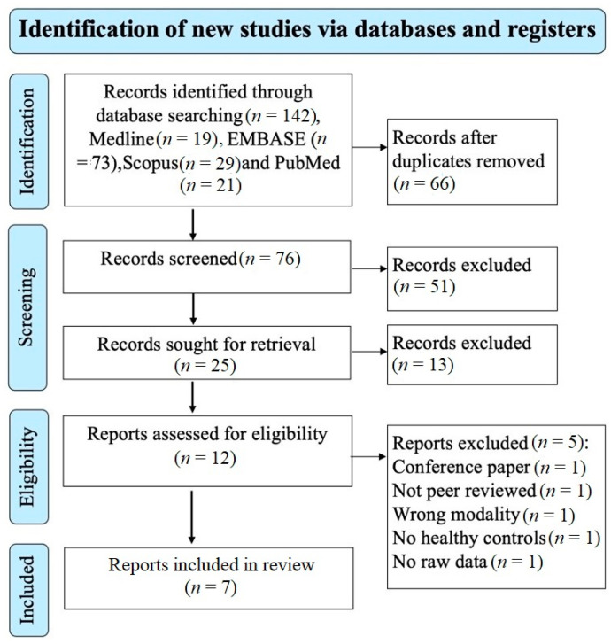
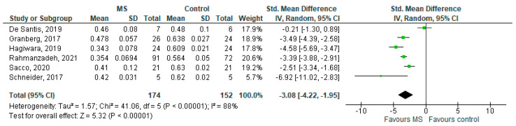
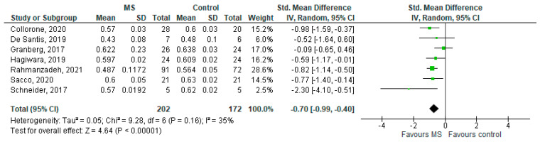
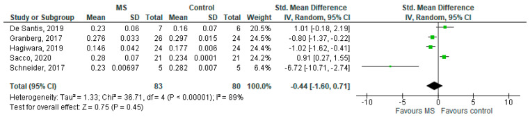
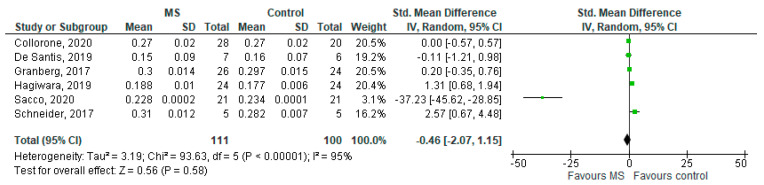
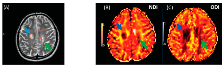
Multiple sclerosis (MS) is characterised by widespread damage of the central nervous system that includes alterations in normal-appearing white matter (NAWM) and demyelinating white matter (WM) lesions. Neurite orientation dispersion and density imaging (NODDI) has been proposed to provide a precise characterisation of WM microstructures. NODDI maps can be calculated for the Neurite Density Index (NDI) and Orientation Dispersion Index (ODI), which estimate orientation dispersion and neurite density. Although NODDI has not been widely applied in MS, this technique is promising in investigating the complexity of MS pathology, as it is more specific than diffusion tensor imaging (DTI) in capturing microstructural alterations. We conducted a meta-analysis of studies using NODDI metrics to assess brain microstructural changes and neuroaxonal pathology in WM lesions and NAWM in patients with MS. Three reviewers conducted a literature search of four electronic databases. We performed a random-effect meta-analysis and the extent of between-study heterogeneity was assessed with the I2 statistic. Funnel plots and Egger's tests were used to assess publication bias. We identified seven studies analysing 374 participants (202 MS and 172 controls). The NDI in WM lesions and NAWM were significantly reduced compared to healthy WM and the standardised mean difference of each was -3.08 (95%CI -4.22 to (-1.95), p ≤ 0.00001, I2 = 88%) and -0.70 (95%CI -0.99 to (-0.40), p ≤ 0.00001, I2 = 35%), respectively. There was no statistically significant difference of the ODI in MS WM lesions and NAWM compared to healthy controls. This systematic review and meta-analysis confirmed that the NDI is significantly reduced in MS lesions and NAWM than in WM from healthy participants, corresponding to reduced intracellular signal fraction, which may reflect underlying damage or loss of neurites.
Keywords: MS; NODDI; multi-compartment diffusion; multi-shell diffusion; multiple sclerosis; neurite orientation dispersion and density imaging.
Conflict of interest statement
The authors declare no conflict of interest.
Figures
References
-
- Filippi M., Rocca M.A., Ciccarelli O., De Stefano N., Evangelou N., Kappos L., Rovira A., Sastre-Garriga J., Tintorè M., Frederiksen J.L., et al. MRI criteria for the diagnosis of multiple sclerosis: MAGNIMS consensus guidelines. Lancet Neurol. 2016;15:292–303. doi: 10.1016/S1474-4422(15)00393-2. - DOI - PMC - PubMed
-
- Wattjes M.P., Rovira À., Miller D., Yousry T.A., Sormani M.P., De Stefano N., Tintoré M., Auger C., Tur C., Filippi M., et al. Evidence-based guidelines: MAGNIMS consensus guidelines on the use of MRI in multiple sclerosis-Establishing disease prognosis and monitoring patients. Nat. Rev. Neurol. 2015;11:597–606. - PubMed
-
- De Groot C.J.A., Bergers E., Kamphorst W., Ravid R., Polman C.H., Barkhof F., Van Der Valk P. Post-mortem MRI-guided sampling of multiple sclerosis brain lesions: Increased yield of active demyelinating and (p)reactive lesions. Brain. 2001;124:1635–1645. doi: 10.1093/brain/124.8.1635. - DOI - PubMed
Publication types
LinkOut - more resources
Full Text Sources
