Case Reports
. 2021 Aug 24;12(9):1295.
doi: 10.3390/genes12091295.

Pathogenic DNM1L Variant (1085G>A) Linked to Infantile Progressive Neurological Disorder: Evidence of Maternal Transmission by Germline Mosaicism and Influence of a Contemporary in cis Variant (1535T>C)

Affiliations
Case Reports

Pathogenic DNM1L Variant (1085G>A) Linked to Infantile Progressive Neurological Disorder: Evidence of Maternal Transmission by Germline Mosaicism and Influence of a Contemporary in cis Variant (1535T>C)

Claudia Piccoli et al. Genes (Basel). .

Abstract

Mitochondria are dynamic organelles undergoing continuous fusion and fission with Drp1, encoded by the DNM1L gene, required for mitochondrial fragmentation. DNM1L dominant pathogenic variants lead to progressive neurological disorders with early exitus. Herein we report on the case of a boy affected by epileptic encephalopathy carrying two heterozygous variants (in cis) of the DNM1L gene: a pathogenic variant (PV) c.1085G>A (p.Gly362Asp) accompanied with a variant of unknown significance (VUS) c.1535T>C (p.Ile512Thr). Amplicon sequencing of the mother's DNA revealed the presence of the PV and VUS in 5% of cells, with the remaining cells presenting only VUS. Functional investigations performed on the patient and his mother's cells unveiled altered mitochondrial respiratory chain activities, network architecture and Ca2+ homeostasis as compared with healthy unrelated subjects' samples. Modelling Drp1 harbouring the two variants, separately or in combination, resulted in structural changes as compared with Wt protein. Considering the clinical history of the mother, PV transmission by a maternal germline mosaicism mechanism is proposed. Altered Drp1 function leads to changes in the mitochondrial structure and bioenergetics as well as in Ca2+ homeostasis. The novel VUS might be a modifier that synergistically worsens the phenotype when associated with the PV.

Keywords: DNM1L; Drp1; encephalopathy; genetic mosaicism; mitochondrial fission.


Conflict of interest statement

The authors declare no conflict of interest.

Figures
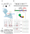
Figure 1
Figure 1
Mutational analysis of DNM1L. (A) Pedigree of the patient family. P*, patient; PM*, patient’s mother; PF*, patient’s father; PAS*, patient’s aborted sibling. Subjects whose biological samples were made available for this study are shown; the smaller square refers to a third conceived by PM deliberately aborted. (B) From the top down: positioning of the DNM1L gene on chromosome 12; gene structure with the two indicated nucleotide variants object of this study; protein domains in the Drp1 primary structure and their schematic assembly in the protein folded structure along with the backbone structure of Dpr1 (left side) modelled using the coordinates of the PDB ID: 3W6O and shown with the indicated domains and location of the variant residues (coloured space-filled rendering); electropherograms showing the DNM1L variants of PBMC of the patient (P) and patient’s mother (PM’), in the saliva of the patient’s mother (PM’’) and in the chorionic villi fibroblast of the patient’s aborted sibling (PAS); enlargements of the PM’ and PM’’ electropherograms are shown at the side. (C) Immunoblotting of PBMC protein extract against anti-Drp1 and β-actin: P, PM as in panel (B); PF, patient’s father; C1, C2, C3 unrelated healthy controls; the figures below each lane are the averaged densitometric values (±SEM) normalized to P from three independent assays.
Figure 2
Figure 2
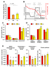
Analysis of the metabolic phenotype in PBMCs. (A) Plasma lactate content in patient (P), patient’s mother (PM) and the averaged value of four independent healthy controls (Cs). (B) Scheme showing the protocol utilized to assay mitochondrial respiratory activity in intact PBMCs cells by high resolution oxymetry; red trace, changes of oxygen content in the assay buffer under baseline and following sequential addition of oligomycin, FCCP and rotenone plus antimycin A; black trace, corresponding oxygen consumption rates (OCRs). (C) OCR/cell number under basal condition, in the presence of oligomycin (proton leak) and in the presence of FCCP (maximum respiration); the reported value are means ± SEM of three independent replicates and are corrected for the rotenone plus antimycin A-insensitive OCR; * P < 0.05 vs. P. (D) Normalized OCRs calculated from the data reported in panel (C); ** P < 0.01. (E) Activities of the respiratory chain complexes I, III and IV and citrate synthase in PBMC cell lysates; the activities were assessed by spectrophotometric assay and the reported values are means ± SEM of three independent replicates; * P < 0.05 vs. P. See Materials and Methods and Results for further details.
Figure 3
Figure 3
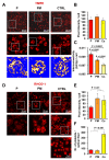
Morpho-functional analysis of the mitochondrial compartment. (A) Confocal microscopy imaging of mtΔΨ assessed by TMRE in PBMCs from patient (P), patient’s mother (PM) and a representative healthy subject (CTRL); the tripartite images show representative images and progressive digital magnifications of the squared details; images at the bottom are rendered as false colour. (B) Quantitative analysis of the TMRE fluorescence/cell. (C) Morphometric evaluation of the TMRE signal/cell with indicated statistical analysis. (D) Confocal microscopy imaging of mitochondrial Ca2+ in PBMCs assessed by Rhod-1; the tripartite images show representative images and digital magnifications of the squared details, and in the CTRL sample a further magnification is reported to highlight the platelets. (E) Histogram showing quantification of the Rhod-1 fluorescence intensity/cell. (F) Histogram showing quantification of the platelet count/optical field. In (B,C,E,F) at least five different optical field/samples, each containing 40–50 cells, were examined and the values averaged ± SD from three independent biological preparations with statistical indication when significant.
Figure 4
Figure 4
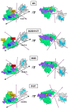
Modelling of the monomeric structure of Wt Drp1 and 362D, 512T variants. The mixed rendered structures were modelled using the coordinates of the PDB ID: 3W6O; two projections rotated by 180° on the X axis are shown. Light blue, residues contributing to the GTP binding site; violet, middle domain (aa 344–489); light green, GSK3β-interaction domain (aa 448–685); blue, homodimerization domain (aa 654–668). The tagged Wt residues 362G and 512I are arrowed in orange and white, respectively; the variants 362D and 512T are arrowed in red and yellow, respectively.
Figure 5
Figure 5
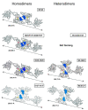
Modelling of the dimeric structure of Wt Drp1 and 362D, 512T variants. The mixed rendered dimeric structures were modelled starting from the monomeric coordinates of the PDB ID: 3W6O. The modelled dimers were constructed using identical monomers (homodimers) or a combination of a Wt monomer and a mutant monomer carrying one or the other of the single variants or a combination of both (heterodimers). The domain essential for dimerization is shown in blue (aa 654–668); residues at position 362 (G or D) and 512 (I or T) are show in red and yellow, respectively.
Figure 6
Figure 6
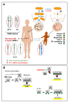
Schematic representation of the proposed pathogenic mechanism linked to the Drp1 variants that are the object of the present study. (A) Model of the mosaicism-based hereditary (AD—autosomic dominant) transmission of the pathogenic variant (PV) and VUS DNM1L in cis variants; mosaicism refers to the presence in an individual of genotipycally distint cells derived from a single zygote (post zygotic event). (B) Model of the impact of the Drp1 variants on the dimer conformations equilibrium and oligomeric assembly and binding to the outer mitochondrial membrane (OMM). See Discussion for further explanation.

References

    1. Westermann B. Mitochondrial fusion and fission in cell life and death. Nat. Rev. Mol. Cell Biol. 2010;11:872–884. doi: 10.1038/nrm3013. - DOI - PubMed
    1. Pernas L., Scorrano L. Mito-Morphosis: Mitochondrial Fusion, Fission, and Cristae Remodeling as Key Mediators of Cellular Function. Annu. Rev. Physiol. 2016;78:505–531. doi: 10.1146/annurev-physiol-021115-105011. - DOI - PubMed
    1. Dorn G.W., 2nd Evolving Concepts of Mitochondrial Dynamics. Annu. Rev. Physiol. 2019;81:1–17. doi: 10.1146/annurev-physiol-020518-114358. - DOI - PubMed
    1. Kraus F., Roy K., Pucadyil T.J., Ryan M.T. Function and regulation of the divisome for mitochondrial fission. Nature. 2021;590:57–66. doi: 10.1038/s41586-021-03214-x. - DOI - PubMed
    1. Chan D.C. Mitochondrial Dynamics and Its Involvement in Disease. Annu. Rev. Pathol. 2020;15:235–259. doi: 10.1146/annurev-pathmechdis-012419-032711. - DOI - PubMed

Publication types

MeSH terms