. 2021 Aug 28;11(9):2531.
doi: 10.3390/ani11092531.

Maternal Under- and Over-Nutrition during Gestation Causes Islet Hypertrophy and Sex-Specific Changes to Pancreas DNA Methylation in Fetal Sheep

Affiliations

Maternal Under- and Over-Nutrition during Gestation Causes Islet Hypertrophy and Sex-Specific Changes to Pancreas DNA Methylation in Fetal Sheep

Maria Peterson et al. Animals (Basel). .

Abstract

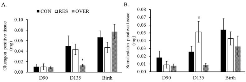
The mechanisms by which fetal programming predisposes offspring to reduced β-cell function later in life are poorly understood. We hypothesized that maternal under- and over-nutrition during gestation would negatively affect offspring pancreas development and alter DNA methylation patterns. Pregnant ewes (n = 78) were fed 100, 60, or 140% of NRC requirements beginning at d 30.2 ± 0.2 of gestation. The fetuses are referred to as CON, RES, and OVER, respectively. Fetal pancreas tissue was collected at d 90 or 135 of gestation or within 24 h of birth. Tissue was preserved for histological (n = 8 to 9 offspring per treatment per time point) and DNA methylation analyses (n = 3 to 4 fetuses per treatment per sex). At d 135, OVER exhibited an increased islet size, reduced islet number, and greater insulin positive area compared with CON (p ≤ 0.03). An increased islet size was also observed at d 135 in RES (p ≤ 0.03) compared with CON. Cellular proliferation was reduced at birth in OVER vs. CON (p = 0.01). In the RES vs. CON females, 62% of the differentially methylated regions (DMRs) were hypomethylated (p ≤ 0.001). In the RES vs. CON males, 93% of the DMRs were hypermethylated (p ≤ 0.001). In OVER, 66 and 80% of the DMRs were hypermethylated in the female and male offspring compared with CON (p ≤ 0.001). In conclusion, changes to maternal diet during pregnancy affects the islet hypertrophy and cellular proliferation of the offspring at early post-natal time points. Additionally, changes in DNA methylation patterns appear to be in a diet-specific and sex-dependent manner.

Keywords: DNA methylation; beta cell function; endocrine pancreas; fetal programming; sheep.


Conflict of interest statement

The authors declare no conflict of interest. The funders had no role in the design of the study; in the collection, analyses, or interpretation of data; in the writing of the manuscript, or in the decision to publish the results.

Figures

Figure 1
Figure 1
Alterations to maternal diet increase fetal pancreas islet size and reduces islet number. Nuclei number (A), Islet size (B) and number (C) were quantified using ImageJ. Pancreas islets from fetuses were stained with hematoxylin and eosin and imaged at 40× magnification. * p ≤ 0.05 compared with CON.
Figure 2
Figure 2
Maternal over-nutrition increases insulin positive area in the pancreas of fetuses at d 135 of gestation and reduces cellular proliferation at birth. Insulin positive area (A), percent PHH3/Insulin positive nuclei (B) and PHH3 positive nuclei (C) were quantified and false colored using ImageJ. Data were normalized using the mass of the pancreas or total number of nuclei where applicable. * p ≤ 0.05, # 0.05 < p ≤ 0.10 compared with CON.
Figure 3
Figure 3
Maternal over-nutrition reduces glucagon positive area and maternal under-nutrition increases somatostatin positive tissue in the pancreas of offspring. Glucagon positive (A) and somatostatin positive tissue (B) were quantified, and images were false colored using ImageJ. Data were normalized using pancreas mass. * p ≤ 0.05, # 0.05 < p ≤ 0.10 compared with CON.
Figure 4
Figure 4
Differentially methylated region (DMR) locations. A total number of 34, 64, 48, and 25 DMRs were identified for CON vs. RES females, CON vs. RES males, CON vs. OVER females, and CON vs. OVER males, respectively. Locations of DMRs were determined using Ensemble. DMRs were considered to be located in a promoter region if they were within 5 kb upstream from the start site of a gene.
Figure 5
Figure 5
Exposure to nutrient restriction during gestation changes fetal pancreas tissue DNA methylation in a sex-specific manner. Fetuses collected from control- and restricted-fed ewes at d 135 of gestation are referred to as CON and RES, respectively. Blue = hypermethylated; red = hypomethylated. Regions were considered differentially methylated when p ≤ 0.001 and ∆ = 0.10.
Figure 6
Figure 6
Maternal over-nutrition during gestation increases DNA methylation in male and female offspring. Fetuses collected from control- and over-fed ewes at d 135 of gestation are referred to as CON and OVER, respectively. Blue = hypermethylated; red = hypomethylated. Regions were considered differentially methylated when p ≤ 0.001 and ∆ = 0.10.

Similar articles

Cited by

References

    1. Cerf M.E., Williams K., Chapman C.S., Louw J. Compromised-Cell Development and Cell Dysfunction in Weanling Offspring from Dams Maintained on a High-Fat Diet during Gestation. Pancreas. 2007;34:347–353. doi: 10.1097/MPA.0b013e31802ee9ae. - DOI - PubMed
    1. Gittes G.K. Developmental biology of the pancreas: A comprehensive review. Dev. Biol. 2009;326:4–35. doi: 10.1016/j.ydbio.2008.10.024. - DOI - PubMed
    1. Cole L., Anderson M., Antin P.B., Limesand S.W. One process for pancreatic beta-cell coalescence into islets involves an epithelial-mesenchymal transition. J. Endocrinol. 2009;203:19–31. doi: 10.1677/JOE-09-0072. - DOI - PMC - PubMed
    1. Wilson M.E., Scheel D., German M.S. Gene expression cascades in pancreatic development. Mech. Dev. 2003;120:65–80. doi: 10.1016/S0925-4773(02)00333-7. - DOI - PubMed
    1. Wu G., Bazer F.W., Wallace J.M., Spencer T.E. Board-invited review: Intrauterine growth retardation: Implications for the animal sciences. J. Anim. Sci. 2006;84:2316–2337. doi: 10.2527/jas.2006-156. - DOI - PubMed

LinkOut - more resources