. 2021 Sep 21;11(9):2755.
doi: 10.3390/ani11092755.

Some Aspects of Development and Histological Structure of the Visual System of Nothobranchius Guentheri

Affiliations

Some Aspects of Development and Histological Structure of the Visual System of Nothobranchius Guentheri

Dmitry L Nikiforov-Nikishin et al. Animals (Basel). .

Abstract

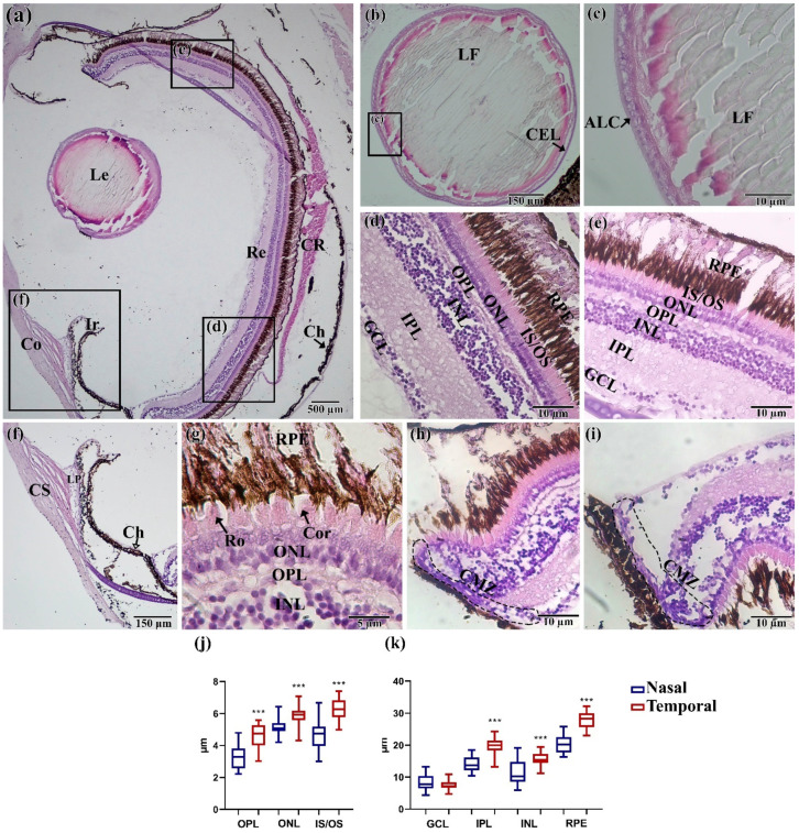
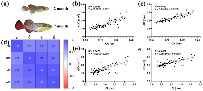
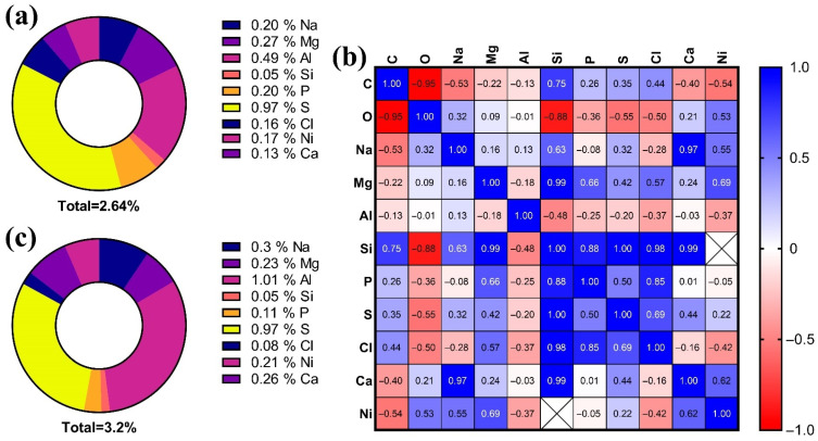
In this, work some aspects of the development of the visual system of Nothobranchius guentheri at the main stages of ontogenesis were described for the first time. It was possible to establish that the formation of the visual system occurs similarly to other representatives of the order Cyprinodontiformes, but significantly differs in terms of the individual stages of embryogenesis due to the presence of diapause. In the postembryonic period, there is a further increase in the size of the fish's eyes and head, to the proportions characteristic of adult fish. The histological structure of the eye in adult N. guentheri practically does not differ from most teleost fish living in the same environmental conditions. The study of the structure of the retina showed the heterogeneity of the thickness of the temporal and nasal areas, which indicates the predominant role of peripheral vision. Morphoanatomical measurements of the body and eyes of N. guentheri showed that their correlation was conservative. This indicates an important role of the visual system for the survival of fish in natural conditions, both for the young and adults. In individuals of the older age group, a decrease in the amount of sodium (Na) and an increase in magnesium (Mg) and calcium (Ca) were found in the eye lens. Such changes in the elemental composition of the lens can be a sign of the initial stage of cataractogenesis and disturbances in the metabolism of lens fibers as a result of aging. This allows us to propose N. guentheri as a model for studying the structure, formation, and aging of the visual and nervous systems.

Keywords: elemental composition; embryogenesis; evolutionary aspects; killifish; lens; morphology; morphometry.


Conflict of interest statement

The authors declare no conflict of interest.

Figures

Figure 1
Figure 1
Morphological characteristics measured in this work. Sl—standard length; lCA—head length; dO—eye diameter; sdO—eye area.
Figure 2
Figure 2
Embryonic development of the visual system of N. guentheri in the period from 26 hpf to 21 dpf: (a,b) 26 to 28 hpf, early neurula, ocular vesicle formation; (c,d) 96 hpf to 7 dpf early somitogenesis (6–12 somites); (e) 10 to 12 dpf retina pigmentation (30–35 somites); (f) 21-n dpf stage before hatching (duration depends on the course of diapause). Yo—yolk; Ov—optic (eye) vesicle; FB—forebrain; L—lens; MB—mid-brain; PM—Periocular Mesenchyme; SC—sclera; CO—cornea; IR—iris.
Figure 3
Figure 3
Postembryonic development of the visual system of N. guentheri: (a,b) 5 to 10 hph larva; (c,d) 7 dph larva. Ch—choroid; Me—melanophores; L—lens; Co—cornea, Ir—iris.
Figure 4
Figure 4
Histological structure of the eye of N. guentheri: (a) Total cut of the eye; (b,c) lens; (d) temporal part of the retina; (e) nasal part of the retina; (f) cornea and iris; (g) layer of receptor cell; (h,i) ciliary marginal zone; (j,k) thickness of layers in the temporal and nasal areas of the retina (n = 3 × 50). The value (***—p < 0.001) from one-way ANOVA with comparison using Tukey’s post hoc analysis. Le—lens, Re—retina, Co—cornea, Ir—iris, CR—choroid rete, CS—corneal stroma, Ch—choroid, GCL—ganglion cell layer, IPL—inner plexiform layer, INL—inner nuclear layer, OPL—outer plexiform layer, ONL—outer nuclear layer, IS/OS—inner segment/outer segment of the photoreceptor cells, RPE—retinal pigment epithelium/cells, LF—lens fibers, ALC—acellular lens capsule, CEL—cuboidal epithelial lens cells; LP—ligamentum pectinatum; Ro—rods; Con—cones; CMZ—ciliary marginal zone.
Figure 5
Figure 5
Morphoanatomical parameters of N. guentheri adults: (a) N. guentheri of different ages, two and seven months; (b) linear regression ICA and sdO; (c) linear regression ICA and dO; (d) correlation matrix of morphometric parameters; (e) SI and sdO linear regression; (f) SI and dO linear regression.
Figure 6
Figure 6
Elemental composition of the lens of N. guentheri: (a,b) pie diagrams of the elemental composition of the lens, excluding carbon and oxygen, for individuals two and seven months old, respectively; (c) correlation matrix between different elements.

References

    1. De Busserolles F., Fitzpatrick J.L., Paxton J.R., Marshall N.J., Collin S.P. Eye-Size Variability in Deep-Sea Lanternfishes (Myctophidae): An Ecological and Phylogenetic Study. PLoS ONE. 2013;8:e58519. doi: 10.1371/journal.pone.0058519. - DOI - PMC - PubMed
    1. Seritrakul P., Gross J.M. Genetic and epigenetic control of retinal development in zebrafish. Curr. Opin. Neurobiol. 2019;59:120–127. doi: 10.1016/j.conb.2019.05.008. - DOI - PMC - PubMed
    1. Demir N. Ichthyology (Ihtiyoloji) Nobel Publishers; Ankara, Turkey: 2009. p. 252.
    1. Furness A.I. The evolution of an annual life cycle in killifish: Adaptation to ephemeral aquatic environments through embryonic diapause. Biol. Rev. 2016;91:796–812. doi: 10.1111/brv.12194. - DOI - PubMed
    1. Lucas-Sánchez A., Almaida-Pagán P.F., Mendiola P., de Costa J. Nothobranchius as a model for aging studies. A review. Aging Dis. 2014;5:281. - PMC - PubMed

LinkOut - more resources