Review
. 2021 Aug 28;11(9):1559.
doi: 10.3390/diagnostics11091559.

Hemodynamic Monitoring in Sepsis-A Conceptual Framework of Macro- and Microcirculatory Alterations

Affiliations
Review

Hemodynamic Monitoring in Sepsis-A Conceptual Framework of Macro- and Microcirculatory Alterations

Liana Valeanu et al. Diagnostics (Basel). .

Abstract

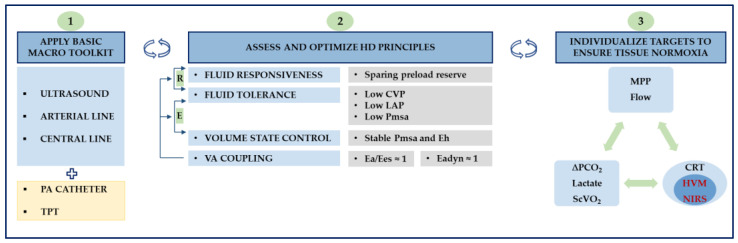
Circulatory failure in sepsis is common and places a considerable burden on healthcare systems. It is associated with an increased likelihood of mortality, and timely recognition is a prerequisite to ensure optimum results. While there is consensus that aggressive source control, adequate antimicrobial therapy and hemodynamic management constitute crucial determinants of outcome, discussion remains about the best way to achieve each of these core principles. Sound cardiovascular support rests on tailored fluid resuscitation and vasopressor therapy. To this end, an overarching framework to improve cardiovascular dynamics has been a recurring theme in modern critical care. The object of this review is to examine the nature of one such framework that acknowledges the growing importance of adaptive hemodynamic support combining macro- and microhemodynamic variables to produce adequate tissue perfusion.

Keywords: hemodynamic; macrocirculation; microcirculation; monitoring; sepsis; shock.


Conflict of interest statement

The authors declare no conflict of interest.

Figures

Figure 1
Figure 1
Interpretation of coupled changes in cardiac output and central venous pressure. CO, cardiac output; CVP, central venous pressure. Adapted with permission [37].
Figure 2
Figure 2
Flow chart for analyzing the hemodynamic profile according to De Backer. ScvO2, central venous oxygen saturation; CO, cardiac output; Microcir, microcirculatory; PvaCO2, venous-to-arterial carbon dioxide partial pressure difference. Reproduced with permission [54].
Figure 3
Figure 3
(a) A schematic illustration of superimposed Frank–Starling (black) and Marik–Phillips (solid blue) curves demonstrating the effects of an identical preload challenge (∆x) on SV and EVLW in a preload-dependent (A) and preload-independent state (B). A: a steep increase in SV (SV1 → SV2) with minimal increase in EVLW (EVLW1 → EVLW2); B: a minimal increase in SV (SV2 → SV3) with a steep increase in EVLW (EVLW2 → EVLW3). Sepsis alters the capillary permeability resulting in a leftward shift of the EVLW curve (dotted blue). EVLW, extravascular lung water; SV, stroke volume; ∆x, a specific preload challenge. Adapted with permission [75]. (b) A schematic illustration of how ventricular performance (i.e., global RV/LV efficiency) and volume state (i.e., MSFP) interact to produce either increased CVP resulting in extrathoracic congestion (e.g., liver, kidney, mesenteric), increased LAP resulting in pulmonary edema, or a mixture of both. Increased permeability independently aggravates tissue congestion. Decompartmentalization occurs in severe conditions, resulting in generalized edema. A to B: for the same volume state, decreased RV/LV efficiency risks fluid intolerance. A to C: preserved RV/LV efficiency does not guarantee fluid tolerance with fluid loading. As a corollary, a normal volume state (i.e., MSFP) does not ensure fluid tolerance in case of severely impaired RV/LV efficiency. Increased CVP could also result from an altered Ecw/El ratio (e.g., intra-abdominal hypertension). CVP, central venous pressure; Ecw, chest wall elastance; El, lung elastance; LAP, left atrial pressure; LV, left ventricle; MSFP, mean systemic filling pressure; RV, right ventricle.
Figure 4
Figure 4
A schematic illustration of ventricular–arterial coupling (VAC). The left ventricle (LV) is characterized by the end-systolic and end-diastolic pressure–volume relationship (ESPVR and EDPVR). The end-systolic LV elastance (Ees) is the slope of the ESPVR line. V0, the LV end-systolic unstressed volume, is the intercept of ESPVR with the volume axis. The arterial system is characterized by the arterial elastance (Ea), i.e., the slope of the Ea line that connects the end-diastolic volume (EDV) with the end-systolic point (red dot). End-systolic coordinates are the end-systolic pressure (ESP) and the end-systolic volume (EDV). Stroke work (SW) (blue area) is maximum for Ea/Ees of 1 and a corresponding ejection fraction (EF) of 50%. LV metabolic efficiency (LVeff) is maximum for Ea/Ees close to 0.5 and a corresponding EF of 66%. PE, end-systolic potential energy (green area). Adapted after Hayashida et al. [80].
Figure 5
Figure 5
A schematic illustration of the steady-state interaction (red dot) between venous return (blue curve) and cardiac function (black curve) introducing Parkin’s Guytonian perspective on global heart efficiency. c, anthropometric constant; CO, cardiac output; CVP, central venous pressure; Eh, global heart efficiency; Evol, volume efficiency; MAP, mean arterial pressure; Pmsa, mean systemic filling pressure analogue; RVR, resistance to venous return; VR, venous return; VRdP, pressure gradient for venous return; ∆, change after a preload challenge. See Appendix A, Table A1 for further discussion.
Figure 6
Figure 6
Conceptualized approach to shock management. Panel 1: a minimal hemodynamic toolkit is presented, encompassing ultrasound, arterial and central venous lines. Extended monitoring comprises pulmonary artery (PA) catheter and transpulmonary thermodilution (TPT). Panel 2: during active resuscitation (R), fluid responsiveness and fluid tolerance need simultaneous assessment, ensuring that: (1) preload reserve is spared, and (2) the minimum increase in intravascular pressures required to sustain adequate tissue perfusion is targeted to guard fluid tolerance. As a corollary, deterioration of fluid tolerance is poised to hamper tissue perfusion and hence requires resolution. During evacuation (E), fluid tolerance and volume state control need simultaneous assessment, ensuring that fluid removal rate is tuned to reach efficiency (lower CVP) and tolerance (preserved CO and MAP). Overall, stable Pmsa and Eh guarantee hemodynamic stability during evacuation phases. In preload independent states, higher removal rates and lower Pmsa can be achieved safely until a threshold is reached when further decreasing Pmsa would result in impaired Eh and low CO. VA coupling represents an energetic refinement of the cardiovascular state that may be superimposed regardless of phase. For practical reasons, Ea/Ees and Eadyn are set to 1 (See Table A1 and Table A2 for further discussion). Panel 3: An integrative approach encompassing macro- and microvascular targets is schematized, emphasizing that target individualization is paramount to improving outcomes. From a microcirculatory perspective, bedside clinicians must rely on clinical examination (i.e., CRT) until more objective monitoring (i.e., HVM) becomes available. CO, cardiac output; CRT, capillary refill time; CVP, central venous pressure; Ea, arterial elastance; Eadyn, dynamic arterial elastance; Ees, ventricular elastance; Eh, global heart efficiency; HVM, handheld vital microscope; LAP, left atrial pressure; MAP, mean arterial pressure; MPP, mean perfusion pressure; NIRS, near-infrared spectroscopy; Pmsa, mean systemic filling pressure analogue; ScVO2, central venous oxygen saturation; VA, ventriculoarterial; ∆PCO2, venous-to-arterial carbon dioxide partial pressure difference.

References

    1. Rhodes A., Evans L., Alhazanni W., Levy M., Antonelli M., Ferrer R., Kumar A., Sevransky J., Sprung C.L., Nunnally M.E., et al. Surviving Sepsis Campaign: International Guidelines for Management of Sepsis and Septic Shock: 2016. Crit. Care Med. 2017;45:486–552. doi: 10.1097/CCM.0000000000002255. - DOI - PubMed
    1. Singer M., Deutschman C.S., Seymour C.C., Shankar-Hari M., Annane D., Bauer M., Bellomo R., Bernard G.R., Chiche J.-D., Coopersmith C.C., et al. The Third International Consensus Definitions for Sepsis and Septic Shock (Sepsis-3) JAMA. 2016;315:801–810. doi: 10.1001/jama.2016.0287. - DOI - PMC - PubMed
    1. Hendy A., Bubenek-Turconi Ş.-I. The Diagnosis and Hemodynamic Monitoring of Circulatory Shock: Current and Future Trends. J. Crit. Care Med. 2016;2:115–123. doi: 10.1515/jccm-2016-0018. - DOI - PMC - PubMed
    1. De Backer D., Hollenberg S., Boerma C., Goedhart P., Büchele G., Ospina-Tascon G., Dobbe I., Ince C. How to evaluate the microcirculation: Report of a round table conference. Crit. Care. 2007;11:R101. doi: 10.1186/cc6118. - DOI - PMC - PubMed
    1. Johansson P., Stensballe J., Ostrowski S. Shock induced endotheliopathy (SHINE) in acute critical illness—A unifying pathophysiologic mechanism. Crit. Care. 2017;21:25. doi: 10.1186/s13054-017-1605-5. - DOI - PMC - PubMed

LinkOut - more resources