. 2021 Sep 20;11(9):1726.
doi: 10.3390/diagnostics11091726.

Usefulness of Endoscopic Ultrasound with the Jelly-Filling Method for Esophageal Varices

Affiliations

Usefulness of Endoscopic Ultrasound with the Jelly-Filling Method for Esophageal Varices

Tsunetaka Kato et al. Diagnostics (Basel). .

Abstract

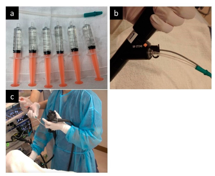
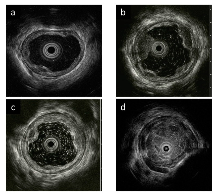
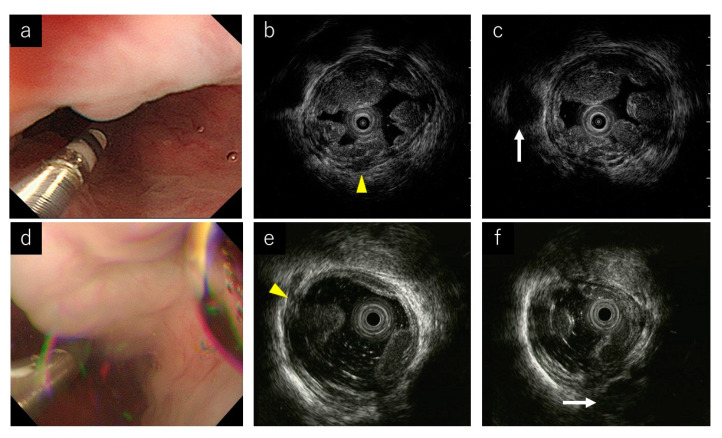
Although the importance of endoscopic ultrasound (EUS) for esophageal varices (EVs) has been demonstrated, it is difficult to obtain sufficient EUS images with the water-filling method because of poor water stagnation in the esophagus. In this study on EVs, we aimed to evaluate the usefulness of the jelly-filling method for EUS. Consecutive patients who underwent EUS for EVs were included. The quality of EUS images, the diagnostic ability of the presence of blood vessels inside and outside the esophageal wall, and the procedure time were compared between the jelly-filling and water-filling methods. Thirty cases were analyzed (jelly-filling method in 13 and water-filling method in 17). The EUS image quality score was significantly higher in the jelly-filling method (jelly vs. water; three points vs. two points, p < 0.001). Additionally, EUS image quality scores in both nonexperts and experts were significantly higher in the jelly-filling method. The diagnostic ability of the presence of perforation veins was significantly higher in the jelly-filling method (jelly vs. water; 100% vs. 52.9%, p = 0.004). However, the procedure time was significantly longer in the jelly-filling method (p = 0.024). In conclusion, EUS using the jelly-filling method for EVs provided sufficient image quality.

Keywords: endoscopic ultrasound; endoscopy; esophageal varices; jelly; portal hypertension.


Conflict of interest statement

The authors declare no conflict of interest.

Figures

Figure 1
Figure 1
The jelly-filling method in the endoscopic ultrasonography (EUS). (a) Jellies is prepared in 5-mL syringes, and a short 14Fr catheter is also prepared. (b) After inserting the endoscope, insert the 14Fr catheter through the wide forceps channel and insert the ultrasound probe through the other forceps channel. (c) During EUS observation, additional jelly injections are performed by the assistant, not the operator.
Figure 2
Figure 2
Evaluation criteria for endoscopic ultrasonography (EUS) image quality. (a) Excellent (4 points). Clear images with no debris-like artifacts in EUS. (b) Good (3 points). A few debris-like artifacts are present, but do not interfere with the evaluation of esophageal varices (EVs) in EUS. (c) Moderate (2 points). Debris-like artifacts are noticeable, but do not interfere with EV evaluation in EUS. (d) Poor (1 point). Difficult to assess EV due to debris-like artifacts and poor esophageal lumen expansion in EUS.
Figure 3
Figure 3
Endoscopic ultrasound (EUS) images of representative cases. (a) Endoscopic image during the jelly-filling method. (b,c) EUS images of the jelly-filling method. Yellow arrow head shows a perforating vein (Pv). White arrow shows a para-esophageal vein (Para-v). (d) Endoscopic image during the water-filling method. (e,f) EUS images of the water-filling method. Yellow arrow head shows a perforating vein (Pv). White arrow shows a para-esophageal vein (Para-v).

References

    1. Jensen D.M. Endoscopic screening for varices in cirrhosis: Findings, implications, and outcomes. Gastroenterology. 2002;122:1620–1630. doi: 10.1053/gast.2002.33419. - DOI - PubMed
    1. Pagliaro L., D’Amico G., Sörensen T.I., Lebrec D., Burroughs A.K., Morabito A., Tiné F., Politi F., Traina M. Prevention of first bleeding in cirrhosis. A meta-analysis of randomized clinical trials of nonsurgical treatment. Ann. Intern. Med. 1992;117:59–70. doi: 10.7326/0003-4819-117-1-59. - DOI - PubMed
    1. D’Amico G., Pagliaro L., Bosch J. The treatment of portal hypertension: A meta-analytic review. Hepatology. 1995;22:332–354. doi: 10.1002/hep.1840220145. - DOI - PubMed
    1. Triantos C., Kalafateli M. Primary prevention of bleeding from esophageal varices in patients with liver cirrhosis. World J. Hepatol. 2014;6:363–369. doi: 10.4254/wjh.v6.i6.363. - DOI - PMC - PubMed
    1. Van Stiegmann G., Cambre T., Sun J.H. A new endoscopic elastic band ligating device. Gastrointest. Endosc. 1986;32:230–233. doi: 10.1016/S0016-5107(86)71815-4. - DOI - PubMed

LinkOut - more resources