. 2021 Aug 27;13(9):1347.
doi: 10.3390/pharmaceutics13091347.

Lung Targeted Lipopolymeric Microspheres of Dexamethasone for the Treatment of ARDS

Affiliations

Lung Targeted Lipopolymeric Microspheres of Dexamethasone for the Treatment of ARDS

Sabna Kotta et al. Pharmaceutics. .

Abstract

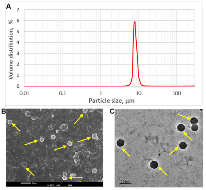
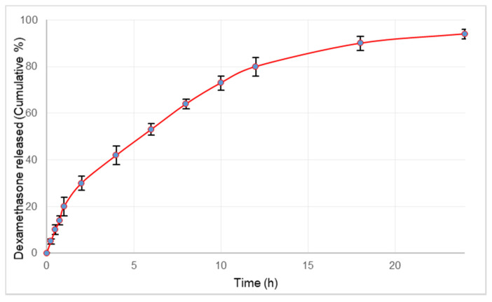
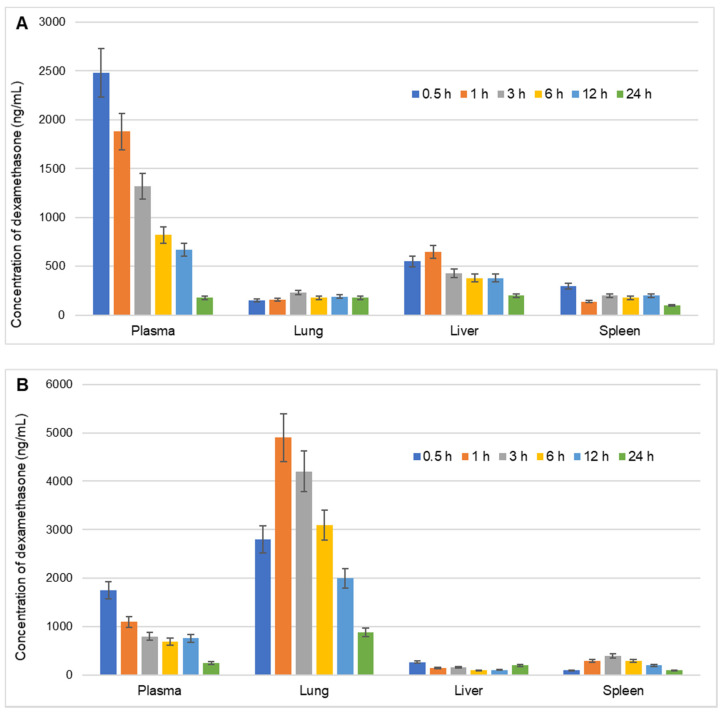
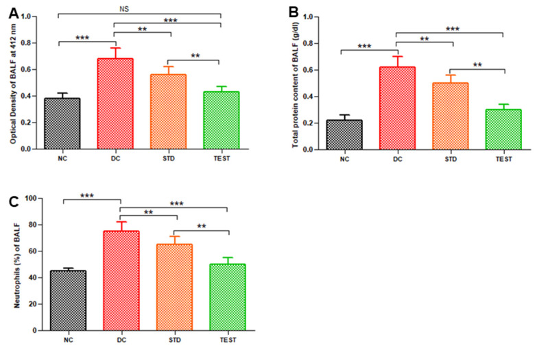
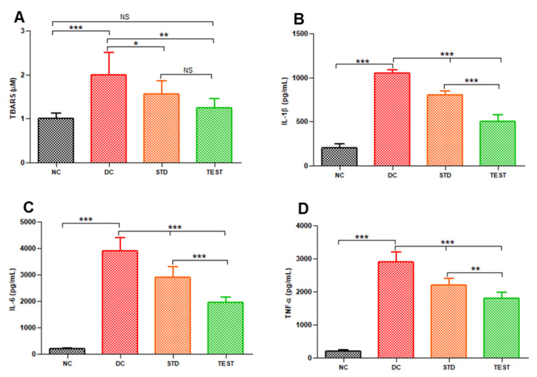
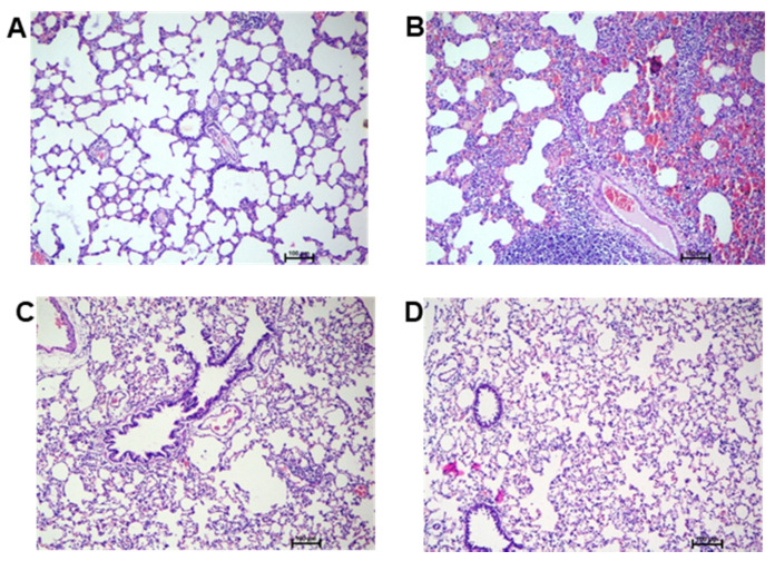
Acute respiratory distress syndrome (ARDS), a catastrophic illness of multifactorial etiology, involves a rapid upsurge in inflammatory cytokines that leads to hypoxemic respiratory failure. Dexamethasone, a synthetic corticosteroid, mitigates the glucocorticoid-receptor-mediated inflammation and accelerates tissue homeostasis towards disease resolution. To minimize non-target organ side effects arising from frequent and chronic use of dexamethasone, we designed biodegradable, lung-targeted microspheres with sustained release profiles. Dexamethasone-loaded lipopolymeric microspheres of PLGA (Poly Lactic-co-Glycolic Acid) and DPPC (Dipalmitoylphosphatidylcholine) stabilized with vitamin E TPGS (D-α-tocopheryl polyethylene glycol succinate) were prepared by a single emulsion technique that had a mean diameter of 8.83 ± 0.32 μm and were spherical in shape as revealed from electron microscopy imaging. Pharmacokinetic and biodistribution patterns studied in the lungs, liver, and spleen of Wistar rats showed high selectivity and targeting efficiency for the lung tissue (re 13.98). As a proof-of-concept, in vivo efficacy of the microspheres was tested in the lipopolysaccharide-induced ARDS model in rats. Inflammation markers such as IL-1β, IL-6, and TNF-α, quantified in the bronchoalveolar lavage fluid indicated major improvement in rats treated with dexamethasone microspheres by intravenous route. Additionally, the microspheres substantially inhibited the protein infiltration, neutrophil accumulation and lipid peroxidation in the lungs of ARDS bearing rats, suggesting a reduction in oxidative stress. Histopathology showed decreased damage to the pulmonary tissue upon treatment with the dexamethasone-loaded microspheres. The multipronged formulation technology approach can thus serve as a potential treatment modality for reducing lung inflammation in ARDS. An improved therapeutic profile would help to reduce the dose, dosing frequency and, eventually, the toxicity.

Keywords: ARDS; dexamethasone; lung inflammation; lung targeting; microspheres.


Conflict of interest statement

The authors declare no conflict of interest.

Figures

Figure 1
Figure 1
Representative graph showing particle size analysis (A), SEM image (B) and TEM image (C) of dexamethasone-loaded microspheres.
Figure 2
Figure 2
Release profile of dexamethasone from microspheres over a period of 24 h. Values plotted are the mean of 6 determinations and error bars indicate standard deviation.
Figure 3
Figure 3
Biodistribution of dexamethasone in various organs namely, liver, lungs, and spleen (ng/g) and plasma (ng/mL) of rats at different time intervals over a period of 24 h after intravenous injection of (A) Conventional marketed injection of dexamethasone and (B) Dexamethasone-loaded lipopolymeric microspheres. Values are the mean of 3 determinations and error bars indicate standard deviation.
Figure 4
Figure 4
Pulmonary haemorrhage in terms of optical density of BALF (A), total protein (B), and neutrophil (C) in BALF from various groups namely, Normal Control (NC), Disease Control (DC), standard treatment of conventional dexamethasone injection (STD), and test formulation treatment with dexamethasone microspheres (TEST) (n = 6). ** and *** represent significant differences at p < 0.01 and p < 0.001, respectively, by Newman–Keuls analysis following ANOVA at 95% confidence limit. NS = Non-significant.
Figure 5
Figure 5
Levels of lipid peroxidation and various inflammatory markers in BALF from various groups namely, Normal Control (NC), Disease Control (DC), standard treatment with conventional dexamethasone injection (STD) and test formulation treatment with dexamethasone microspheres (TEST) (n = 6) (A) TBARS, (B) IL-1β, (C) IL-6, and (D) TNF-α. *, **, and *** represents significant difference at p < 0.05 and p < 0.01 and p < 0.001, respectively, by Newman–Keuls analysis following ANOVA at 95% confidence limit. NS = Not significant.
Figure 6
Figure 6
Haematoxylin and eosin (H,E)-stained representative rat lung tissue histopathology images of (A) normal control, (B) diseased control, (C) standard treatment with conventional dexamethasone injection, and (D) developed test formulation treatment.

References

    1. Marini J.J., Gattinoni L. Management of COVID-19 Respiratory Distress. JAMA. 2020;323:2329–2330. doi: 10.1001/jama.2020.6825. - DOI - PubMed
    1. Kotta S., Aldawsari H.M., Badr-Eldin S.M., Alhakamy N.A., Md S., Nair A.B., Deb P.K. Combating the Pandemic COVID-19: Clinical Trials, Therapies and Perspectives. Front. Mol. Biosci. 2020;7:606393. doi: 10.3389/fmolb.2020.606393. - DOI - PMC - PubMed
    1. Papanikolaou I.C., Tsenempi X.A. Tropical Lung Diseases. Hunt. Trop. Med. Emerg. Infect. Dis. 2020:1–7. doi: 10.1016/B978-0-323-55512-8.00001-6. - DOI
    1. Gibson P.G., Qin L., Puah S.H. COVID-19 acute respiratory distress syndrome (ARDS): Clinical features and differences from typical pre-COVID-19 ARDS. Med. J. Aust. 2020;213:54–56.e51. doi: 10.5694/mja2.50674. - DOI - PMC - PubMed
    1. Máca J., Jor O., Holub M., Sklienka P., Burša F., Burda M., Janout V., Ševčík P. Past and Present ARDS Mortality Rates: A Systematic Review. Respir. Care. 2017;62:113–122. doi: 10.4187/respcare.04716. - DOI - PubMed