

Dual Emissive Ir(III) Complexes for Photodynamic Therapy and Bioimaging
- PMID: 34575458
- PMCID: PMC8472790
- DOI: 10.3390/pharmaceutics13091382
Dual Emissive Ir(III) Complexes for Photodynamic Therapy and Bioimaging
Abstract
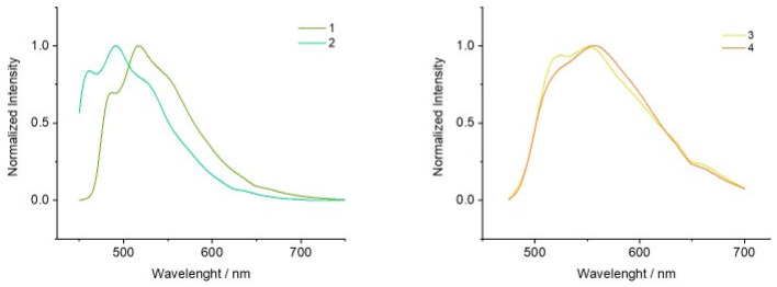
Photodynamic therapy (PDT) is a cancer treatment still bearing enormous prospects of improvement. Within the toolbox of PDT, developing photosensitizers (PSs) that can specifically reach tumor cells and promote the generation of high concentration of reactive oxygen species (ROS) is a constant research goal. Mitochondria is known as a highly appealing target for PSs, thus being able to assess the biodistribution of the PSs prior to its light activation would be crucial for therapeutic maximization. Bifunctional Ir(III) complexes of the type [Ir(C^N)2(N^N-R)]+, where N^C is either phenylpyridine (ppy) or benzoquinoline (bzq), N^N is 2,2'-dipyridylamine (dpa) and R either anthracene (1 and 3) or acridine (2 and 4), have been developed as novel trackable PSs agents. Activation of the tracking or therapeutic function could be achieved specifically by irradiating the complex with a different light wavelength (405 nm vs. 470 nm respectively). Only complex 4 ([Ir(bzq)2(dpa-acr)]+) clearly showed dual emissive pattern, acridine based emission between 407-450 nm vs. Ir(III) based emission between 521 and 547 nm. The sensitivity of A549 lung cancer cells to 4 evidenced the importance of involving the metal center within the activation process of the PS, reaching values of photosensitivity over 110 times higher than in dark conditions. Moreover, complex 4 promoted apoptotic cell death and possibly the paraptotic pathway, as well as higher ROS generation under irradiation than in dark conditions. Complexes 2-4 accumulated in the mitochondria but species 2 and 4 also localizes in other subcellular organelles.
Keywords: cytotoxicity; dual emiter; fluorescence microscopy; iridium; mitochondria; optical properties; photodynamic therapy.
Conflict of interest statement
The authors declare no conflict of interest.
Figures


References
-
- Dąbrowski J.M. Reactive Oxygen Species in Photodynamic Therapy: Mechanisms of Their Generation and Potentiation. Adv. Inorg. Chem. 2017;70:343–394. doi: 10.1016/bs.adioch.2017.03.002. - DOI
-
- McKenzie L.K., Bryant H.E., Weinstein J.A. Transition metal complexes as photosensitisers in one- and two-photon photodynamic therapy. Coord. Chem. Rev. 2019;379:2–29. doi: 10.1016/j.ccr.2018.03.020. - DOI
Grants and funding
LinkOut - more resources
Full Text Sources
