. 2021 Sep 2;13(9):1386.
doi: 10.3390/pharmaceutics13091386.

Nanostructured Lipid Carrier Gel Formulation of Recombinant Human Thrombomodulin Improve Diabetic Wound Healing by Topical Administration

Affiliations

Nanostructured Lipid Carrier Gel Formulation of Recombinant Human Thrombomodulin Improve Diabetic Wound Healing by Topical Administration

Yuan-Shuo Hsueh et al. Pharmaceutics. .

Abstract

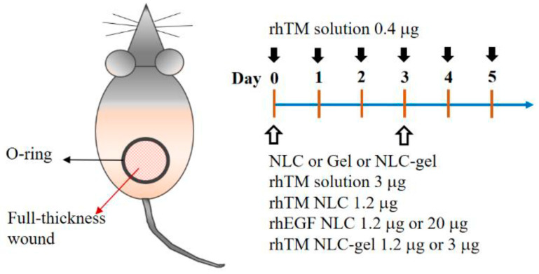
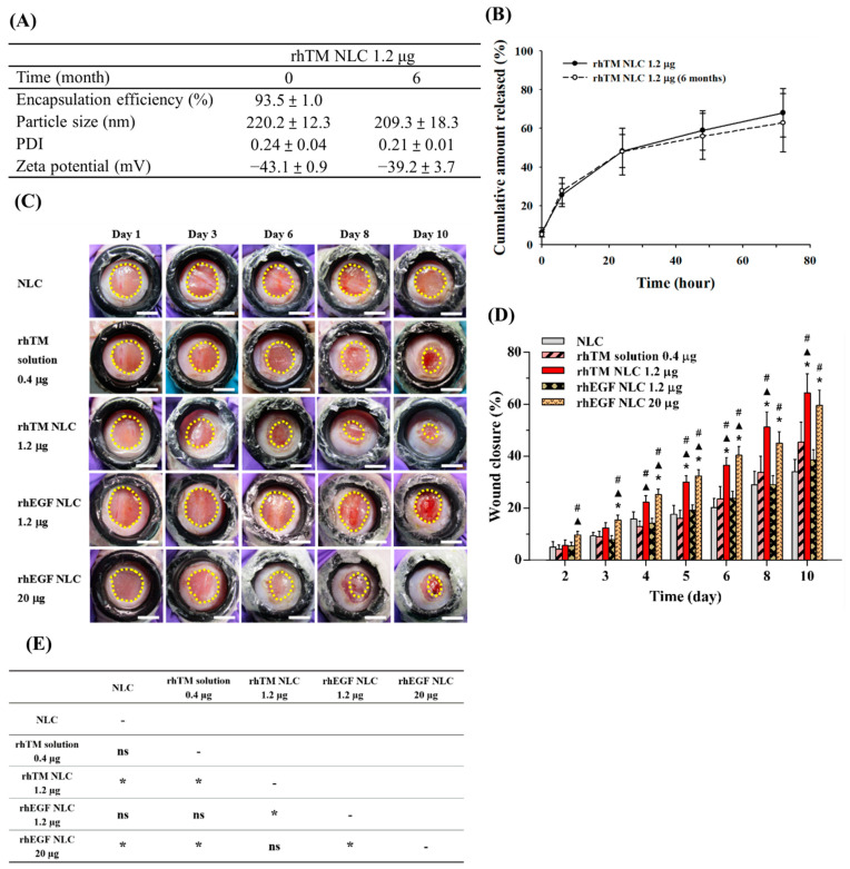
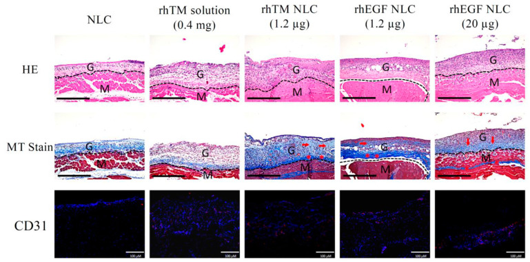
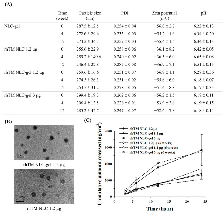
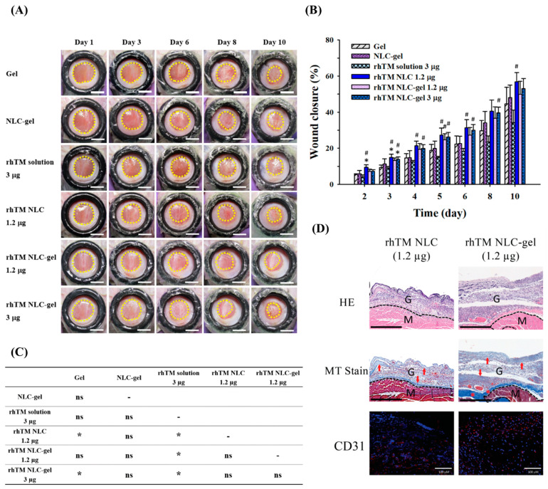
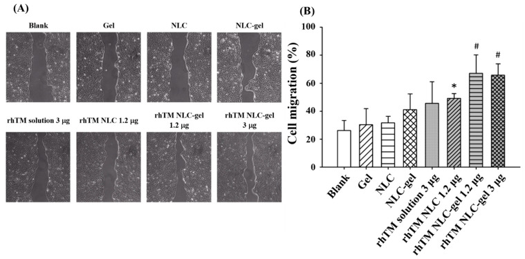
Recombinant human thrombomodulin (rhTM), an angiogenesis factor, has been demonstrated to stimulate cell proliferation, keratinocyte migration and wound healing. The objective of this study was to develop nanostructured lipid carrier (NLC) formulations encapsulating rhTM for promoting chronic wound healing. RhTM-loaded NLCs were prepared and characterized. Encapsulation efficiency was more than 92%. The rate of rhTM release from different NLC formulations was influenced by their lipid compositions and was sustained for more than 72 h. Studies on diabetic mouse wound model suggested that rhTM-NLC 1.2 µg accelerated wound healing and was similar to recombinant human epidermal growth factor-NLC (rhEGF-NLC) 20 µg. By incorporating 0.085% carbopol (a highly crosslinked polyacrylic acid polymer) into rhTM NLC, the NLC-gel presented similar particle characteristics, and demonstrated physical stability, sustained release property and stability within 12 weeks. Both rhTM NLC and rhTM NLC-gel improved wound healing of diabetic mice and cell migration of human epidermal keratinocyte cell line (HaCaT) significantly. In comparison with rhTM solution, plasma concentrations of rhTM post applications of NLC and NLC-gel formulations were lower and more sustained in 24 h. The developed rhTM NLC and rhTM NLC-gel formulations are easy to prepare, stable and convenient to apply to the wound with reduced systemic exposure, which may warrant potential delivery systems for the care of chronic wound patients.

Keywords: angiogenesis factor; carbopol gel; chronic wound healing; nanostructured lipid carrier; protein drug delivery; sustained release; thrombomodulin.


Conflict of interest statement

The authors declare that there is no conflict of interest. The company had no role in the design of the study; in the collection, analyses, or interpretation of data; in the writing of the manuscript, or in the decision to publish the results.

Figures

Figure 1
Figure 1
Topical treatment protocol. The black arrows denoted application days for rhTM solution 0.4 μg. The white arrows indicated application days for NLC, Gel, NLC-gel, rhTM solution 3 μg, rhTM NLC 1.2 μg, rhEGF NLC 1.2 μg, rhEGF NLC 20 μg, rhTM NLC-gel 1.2 μg and rhTM NLC-gel 3 μg, respectively. Each dose was applied in 50 μL solution or gel.
Figure 2
Figure 2
Characterization and wound healing effect of rhTM NLC 1.2 μg. (A) Physical characteristics of rhTM NLC 1.2 μg (n = 3). Data presented as mean ± SD. (B) In vitro drug release of freshly prepared rhTM NLC 1.2 μg and after storage at 4 °C for 6 months. Data presented as mean ± SD. (C) Representative wound area of streptozotocin-induced diabetic mice at Day 1 to 10 with different treatments. (n = 6–15 for each group; Scale bar = 5 mm). (D) Wound closure rate by quantifying wound area and analyzing with mathematical formula described in Methods. Data presented as mean ± SEM. The significance was analyzed using Student’s t test. p < 0.05 compared with NLC group (*), rhTM solution 0.4 µg group (▲), or rhEGF NLC 1.2 µg group (#). (E) Multiple comparison analysis of wound closure effect between different treatment groups based on general linear model. Data presented as p value. Bonferroni post hoc test was applied. (*: p < 0.05; ns: p > 0.05).
Figure 3
Figure 3
RhTM NLC 1.2 μg increased granulation tissues and enhanced collagen deposition and angiogenesis at the wound site. Wounded skins on Day 10 were collected, fixed, and embedded in paraffin. Samples were stained with hematoxylin and eosin (HE), Masson’s trichrome staining (MT), or CD31, respectively. Representative images of central area of wound were shown. In MT staining, arrows pinpointed newly formed collagen (faint blue) and asterisks denoted deposited collagen (deep blue). Dashed lines denoted boundaries between the granulation tissue and dermis/muscle. G, granulation tissue; M, muscle. In CD31 immunostaining, CD31 and nuclei were visualized as red and blue dots, respectively. Scale bar of HE and MT = 400 μm; scale bar of CD31 = 100 μm.
Figure 4
Figure 4
Characteristics and drug release profile of rhTM NLC-gel. (A) Physical characteristics of freshly prepared rhTM NLC formulations with and without gel, and after storage at 4 °C for up to 12 weeks. (n = 3). (B) Morphology of rhTM NLC and rhTM NLC-gel particles under TEM. (Scale bar = 0.2 µm). Data presented as mean ± SD. (C) In vitro drug release profile of freshly prepared (n = 4 each) rhTM NLC and rhTM NLC-gel and after storage at 4 °C for 6 weeks (n = 2 each). Drug release followed dose and time dependent manner.
Figure 5
Figure 5
Wound healing effect of RhTM NLC-gel. (A) Representative wound area of streptozotocin-induced diabetic mice at Day 1 to 10 with different treatments. (n = 5–10 for each group; scale bar = 5 mm) (B) Wound closure of diabetic mice by quantification wound area in (A) and analysis following mathematical formula described in Methods. Data presented as mean ± SEM. The significance was analyzed using Student’s t test. p < 0.05 compared with gel group (*) or rhTM solution 3 µg group (#). (C) Multiple comparison analysis of wound closure effect between different treatment groups based on general linear model. Bonferroni post hoc test was applied. (*: p < 0.05; ns: p > 0.05) (D) Wounded skins on Day 10 were collected, fixed, and embedded in paraffin. The samples were stained with hematoxylin and eosin (HE), Masson’s trichrome staining (MT), or CD31, respectively. Representative images of central area of wound were shown. In MT staining, arrows pinpointed newly formed collagen (faint blue) and asterisks denoted deposited collagen (deep blue). Dashed lines denoted boundaries between the granulation tissue and dermis/muscle. G, granulation tissue; M, muscle. In CD31 immunostaining, CD31 and nuclei were visualized as red and blue dots, respectively. Scale bar of HE and MT = 400 μm; scale bar of CD31 = 100 μm.
Figure 6
Figure 6
RhTM NLC-gel 1.2 μg enhanced cell migration of human keratinocyte HaCaT cells. The cells were seeded, treated with different formulations, and then photographed using OPTIKA camera and Leica microscopy system. (A) Representative images were shown. (B) The ratio of wound recovery was quantitatively determined with ImageJ software. Data are presented as mean ± SD (n = 4 each treatment). The significance was analyzed using Student’s t-test. p < 0.05 compared with Blank/Gel/NLC group (*) or Blank/Gel/NLC/rhTM NLC 1.2 μg group (#).

Similar articles

Cited by

References

    1. Martin C., Low W.L., Amin M.C., Radecka I., Raj P., Kenward K. Current trends in the development of wound dressings, biomaterials and devices. Pharm. Pat. Anal. 2013;2:341–359. doi: 10.4155/ppa.13.18. - DOI - PubMed
    1. Mayet N., Choonara Y.E., Kumar P., Tomar L.K., Tyagi C., Du Toit L.C., Pillay V. A comprehensive review of advanced biopolymeric wound healing systems. J. Pharm. Sci. 2014;103:2211–2230. doi: 10.1002/jps.24068. - DOI - PubMed
    1. Pazyar N., Yaghoobi R., Rafiee E., Mehrabian A., Feily A. Skin wound healing and phytomedicine: A review. Skin Pharmacol. Physiol. 2014;27:303–310. doi: 10.1159/000357477. - DOI - PubMed
    1. Sinno H., Prakash S. Complements and the wound healing cascade: An updated review. Plast. Surg. Int. 2013;2013:146764. doi: 10.1155/2013/146764. - DOI - PMC - PubMed
    1. Wong V.W., Gurtner G.C., Longaker M.T. Wound healing: A paradigm for regeneration. Mayo Clin. Proc. 2013;88:1022–1031. doi: 10.1016/j.mayocp.2013.04.012. - DOI - PubMed

LinkOut - more resources