. 2021 Sep 19;13(9):1518.
doi: 10.3390/pharmaceutics13091518.

Mesoporous Calcium-Silicate Nanoparticles Loaded with Low-Dose Triton-100+Ag+ to Achieve Both Enhanced Antibacterial Properties and Low Cytotoxicity for Dentin Disinfection of Human Teeth

Affiliations

Mesoporous Calcium-Silicate Nanoparticles Loaded with Low-Dose Triton-100+Ag+ to Achieve Both Enhanced Antibacterial Properties and Low Cytotoxicity for Dentin Disinfection of Human Teeth

Mengting Duan et al. Pharmaceutics. .

Abstract

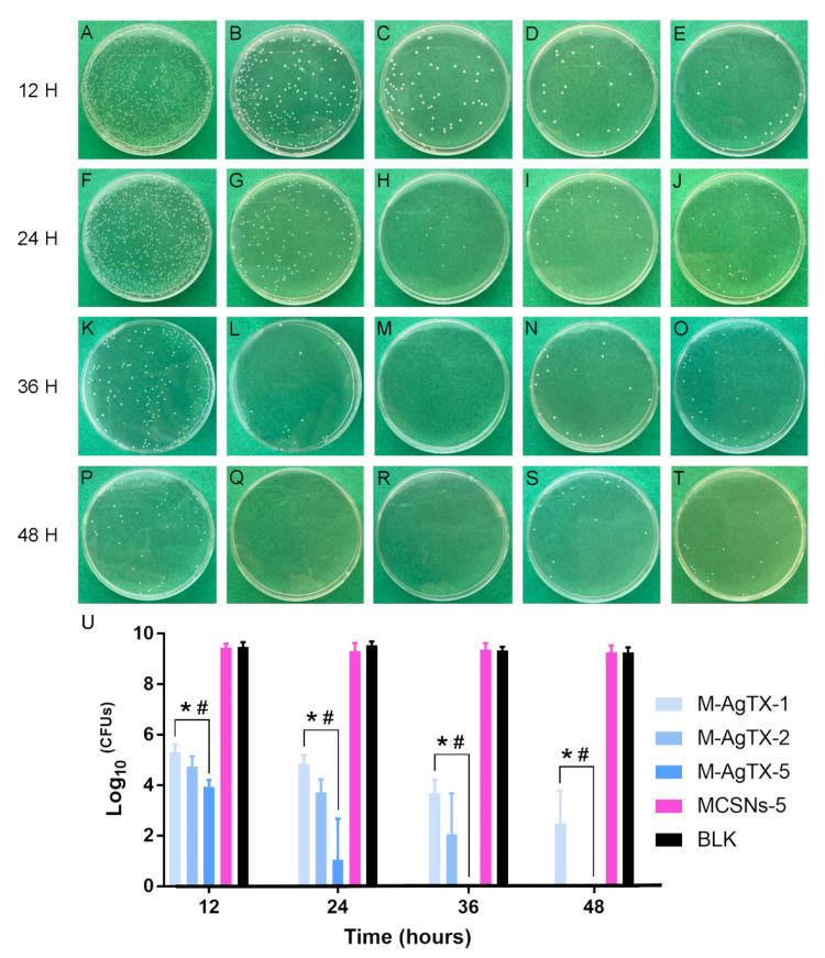
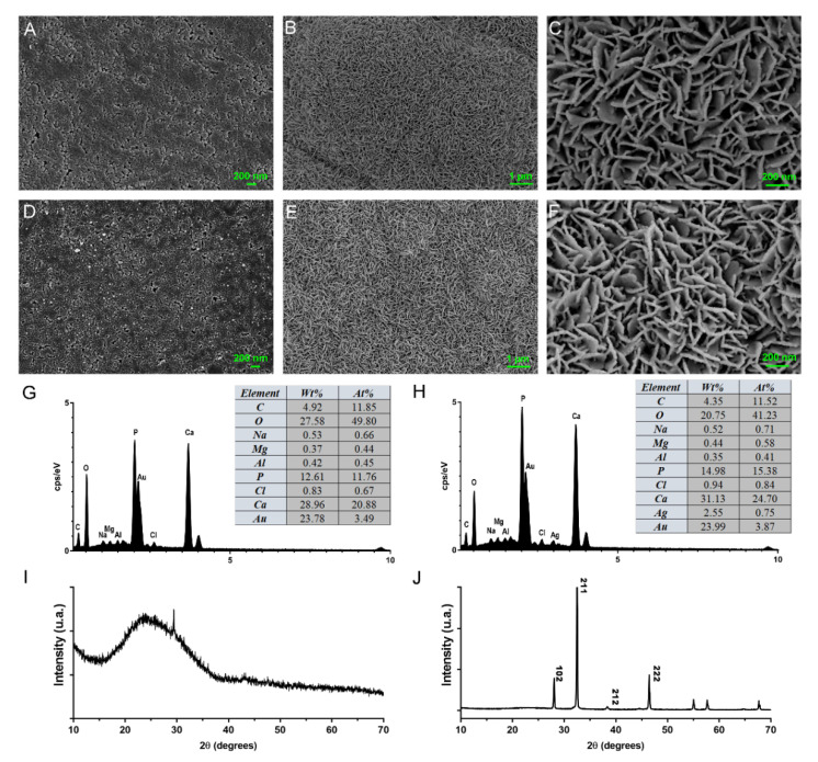
Mesoporous calcium-silicate nanoparticles (MCSNs) are excellent biomaterials for controlled drug delivery and mineralization induction. In this study, MCSNs were loaded with low-dose silver ion (Ag+) and Triton X-100 (TX-100) as the M-AgTX to achieve both enhanced antibacterial properties and low cytotoxicity for dentin disinfection. The physicochemical property, biocompatibility, infiltration ability into dentinal tubules, anti-bacterial ability against both planktonic Enterococcusfaecalis (E. faecalis) and its biofilm on dentin, effects on dentin microhardness and in vitro mineralization property were systematically investigated. Results confirmed that the MCSNs and M-AgTX nanoparticles showed typical morphology of mesoporous materials and exhibited sustained release of chemicals with an alkaline pH value over time. M-AgTX also exhibited excellent biocompatibility on MC3T3-E1 cells and could eliminate 100% planktonic E. faecalis after 48-h treatment. On dentin slices, it could enter dentinal tubules by ultrasonic activation and inhibit the growth of E. faecalis on dentin. M-AgTX could completely inactive 28-day E. faecalis biofilm. TEM confirmed the destruction of cell membrane integrity and Ag+ infiltration into bacteria by M-AgTX. Besides, dentin slices medicated with M-AgTX nanoparticles displayed an increased microhardness. After being immersed in SBF for 7 days, apatite crystals could be observed on the surface of the material tablets. M-AgTX could be developed into a new multifunctional intra-canal medication or bone defect filling material for infected bone defects due to its sustained release profile, low cytotoxicity, infiltration ability, enhanced anti-bacterial and mineralization features.

Keywords: Enterococcus faecalis; Triton X-100; mesoporous; nanoparticle; root canal disinfection; silver.


Conflict of interest statement

The authors declare no conflict of interest.

Figures

Figure 1
Figure 1
Characterization of MCSNs and M-AgTX. (A,D) Representative images of MCSNs and M-AgTX by FE-SEM; (B,E) Representative images of MCSNs and M-AgTX by TEM; (C,F) Representative images of MCSNs and M-AgTX by EDS.
Figure 2
Figure 2
Nitrogen adsorption-desorption isotherm test and pore size distribution of MCSNs and M-AgTX. (A,C) Nitrogen adsorption-desorption isotherm test of MCSNs and M-AgTX, respectively; (B,D) Pore size distribution of MCSNs and M-AgTX, respectively.
Figure 3
Figure 3
FTIR spectrum of MCSNs, M-AgTX and TX-100.
Figure 4
Figure 4
pH and release profile of MCSNs and M-AgTX. (A) pH curve of MCSNs and M-AgTX; (B,C) Total released amount of SiO32−, Ca2+ of MCSNs and M-AgTX, respectively; (D,E) Total released amount of Ag+ and TX-100 of M-AgTX, respectively.
Figure 5
Figure 5
CCK-8 results among groups. (BLK: blank control group; BKG: medium background; M-AgTX-1, M-AgTX-2, M-AgTX-5: 1, 2, 5 mg/mL M-AgTX groups; MCSNs-1, MCSNs-2, MCSNs-5: 1, 2, 5 mg/mL MCSNs groups; *, #: significant difference when compared with 2% CHX group, and BLK group, respectively, p < 0.05.).
Figure 6
Figure 6
Anti-bacterial effects of different concentrations of materials against planktonic E. faecalis at different time intervals. (AC,FH,KM,PR) Representative images of CFUs of 1, 2, 5 mg/mL M-AgTX groups at 12, 24, 36, 48 h; (D,I,N,S) Representative images of CFUs of 5 mg/mL MCSNs group at 12, 24, 36, 48 h after 106 times dilution; (E,J,O,T) Representative images of CFUs of BLK at 12, 24, 36, 48 h after 106 times dilution; (U) Log10 (CFUs) of groups. (BLK: blank control group; M-AgTX-1, M-AgTX-2, M-AgTX-5: 1, 2, 5 mg/mL M-AgTX groups; MCSNs-5: 5 mg/mL MCSNs group; *, #: significant difference when compared with MCSNs-5 group and BLK group, respectively, p < 0.05.).
Figure 7
Figure 7
Infiltration of M-AgTX into dentinal tubules. (A) Original dentinal tubule openings (A) ×5000; (B,C) Tubule openings treated ultrasonically with M-AgTX (B) ×5000, (C) ×20,000); (D) Original dentinal tubule axial cross sections (D) ×5000; (E) Tubule axial cross sections after being treated with M-AgTX (E) ×7000; (F) EDS spectrum of selected square area in (E).
Figure 8
Figure 8
FE-SEM images showing anti-bacterial effects on pretreated dentin slices. (A,D) E. faecalis grown on dentin slice of blank control group (A) ×5000, (D) ×20,000; (B,E) E. faecalis grown on dentin slice of MCSNs pre-treated group (B) ×5000, (E) ×20,000; (C,F) E. faecalis grown on dentin slice of M-AgTX pre-treated group (C) ×5000, (F) ×20,000.
Figure 9
Figure 9
The OD value after the direct soaking of dentin slices from different treatments in fresh BHI media. (A) Pretreated dentin slices; (B) Dentin slices with 4-week E. faecalis biofilm after 7-day medication; (C,D) Comparisons of OD value at 600 nm at 36 h and 48 h of (B). (*,#: significant difference when compared with MSCNs group and BLK group, respectively, p < 0.05.).
Figure 10
Figure 10
FE-SEM images of 4-week E. faecalis biofilm on dentin slices after 7-day medication. (A,B) biofilm treated with PBS gel (A) ×5000, (B) ×20,000; (C,D) biofilm treated with MCSNs paste (C) ×5000, (D) ×20,000); (E,F) biofilm treated with M-AgTX paste (E) ×5000, (F) ×20,000; (G,H) biofilm treated with 2% CHX gel (G) ×5000, (H) ×20,000; biofilm treated with Ca(OH)2 paste (I) ×5000, (J) ×20,000).
Figure 11
Figure 11
TEM images of endocytosis behavior of E. faecalis after being treated with M-AgTX. (AF) Different degree of biological membrane defect. (★ indicating the nanoparticles, arrows in (A) indicating the cell membrane defect, square frame in (D) indicating the M-AgTX outside the cell, and arrow in (E) indicating the normal morphology.
Figure 12
Figure 12
Dentin microhardness measurement. (A) Representative image of dentin slices in microhardness measurement (×40); (B) Comparison of microhardness among groups. (*: p < 0.05).
Figure 13
Figure 13
In vitro mineralization of MCSNs and M-AgTX. (A,D) FE-SEM image of original MCSNs and M-AgTX tablets surfaces, respectively; (B,C) FE-SEM image of MCSNs tablet surface after being soaked in SBF for 7 days (B) ×10,000, (C) ×50,000); (E,F) FE-SEM image of MCSNs tablet surface after being soaked in SBF for 7 days (E) ×10,000, (F) ×50,000; (G,H) EDS spectrum of crystals formed on MCSNs and M-AgTX tablets surfaces after being soaked in SBF for 7 days, respectively; (I,J) Wide-angle XRD patterns of M-AgTX before and after being soaked in SBF for 7 days.

Similar articles

Cited by

References

    1. Figdor D., Davies J.K., Sundqvist G. Starvation survival, growth and recovery of Enterococcus faecalis in human serum. Oral Microbiol. Immunol. 2003;18:234–239. doi: 10.1034/j.1399-302X.2003.00072.x. - DOI - PubMed
    1. Stuart C.H., Schwartz S.A., Beeson T.J., Owatz C.B. Enterococcus faecalis: Its Role in Root Canal Treatment Failure and Current Concepts in Retreatment. J. Endod. 2006;32:93–98. doi: 10.1016/j.joen.2005.10.049. - DOI - PubMed
    1. Carr G.B., Schwartz R.S., Schaudinn C., Gorur A., Costerton J.W. Ultrastructural examination of failed molar retreatment with secondary apical periodontitis: An examination of endodontic biofilms in an endodontic retreatment failure. J. Endod. 2009;35:1303–1309. doi: 10.1016/j.joen.2009.05.035. - DOI - PubMed
    1. Gomes B.P., Vianna M.E., Zaia A.A., Almeida J.F., Souza-Filho F.J., Ferraz C.C. Chlorhexidine in endodontics. Braz. Dent. J. 2013;24:89–102. doi: 10.1590/0103-6440201302188. - DOI - PubMed
    1. Louwakul P., Saelo A., Khemaleelakul S. Efficacy of calcium oxide and calcium hydroxide nanoparticles on the elimination of Enterococcus faecalis in human root dentin. Clin. Oral Investig. 2017;21:865–871. doi: 10.1007/s00784-016-1836-x. - DOI - PubMed

LinkOut - more resources